Design and experimental research of four-channel spherically bent crystal imaging system
-
摘要:
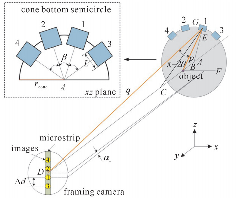
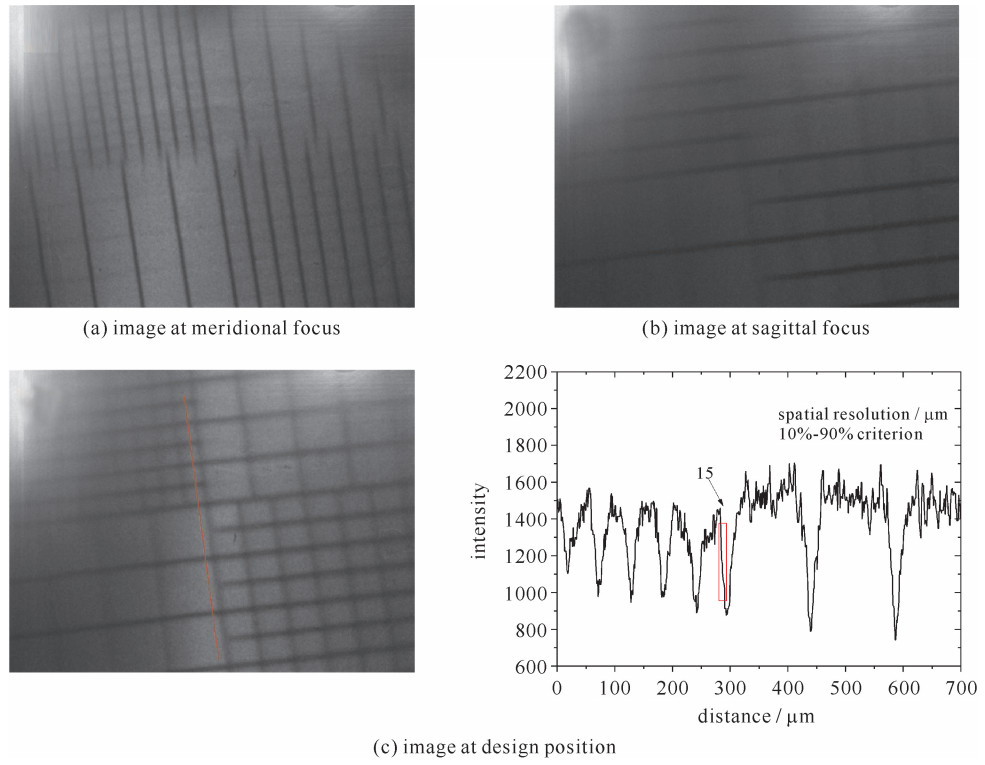
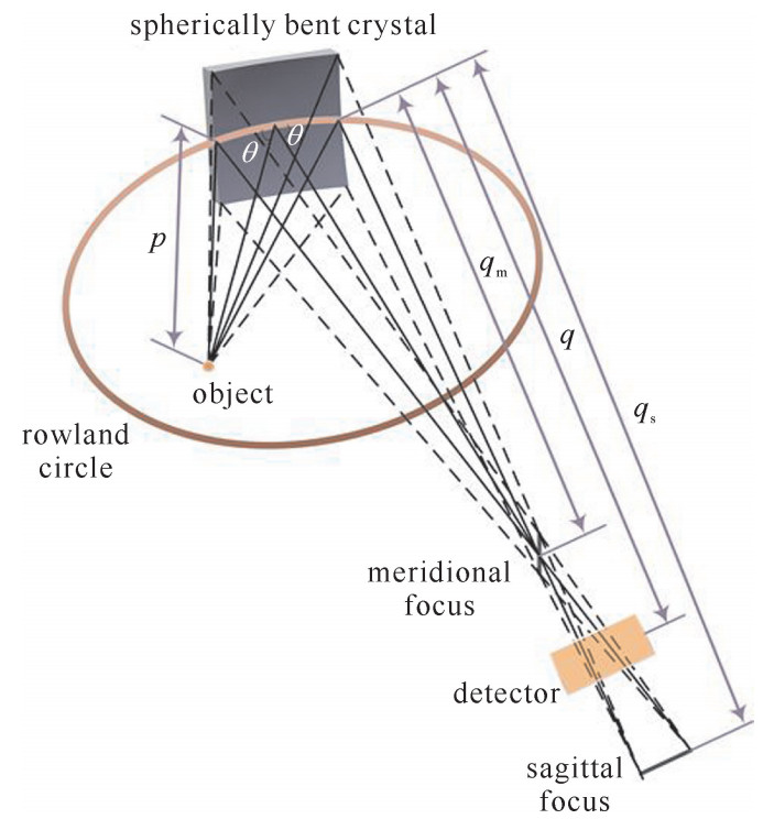
基于动态X射线荧光成像技术对高集光效率、单色化成像诊断设备的需求,提出了一种四通道球面弯晶成像系统设计。采用“圆锥体”空间排布方式,解决了多个通道耦合问题。通过调整弯晶姿态,实现了像点的合理分布。针对4.51 keV能点,采用Ge(400)球面弯晶作为成像元件,给出了四通道弯晶成像系统的光学初始结构参数。在实验中利用Ti靶X射线光管,对单个通道进行了网格背光成像,获得的二维图像放大倍数为7.8倍,空间分辨率达到15 μm,初步验证了系统的成像性能。四通道弯晶成像系统与分幅相机结合,能有效解决动态X射线荧光成像技术信号弱、图像信噪比低的技术难点。
Abstract:Since dynamic X-ray fluorescence imaging technology requires diagnostic equipment which has high throughput and narrow spectral width, we present the design of four-channel spherically bent crystal imaging system. The system adopts a cone spatial configuration to solve the problem of multiple channel coupling. With the size limit of framing camera taken into account, the images are reasonably planned by adjusting the position of bent crystal. We utilize Ge 400 crystal as the imaging component at 4.51 keV and then propose optical initial structural parameters of system. Grid backlit images of single channel are obtained by using X-ray tube in the laboratory. The magnification is 7.8, and the spatial resolution is 15 μm. The results preliminarily verify the performance of system. The four-channel system combined with the framing camera can effectively solve the technical difficulties such as weak signal and low signal to noise ratio in dynamic fluorescence imaging.
-
表 1 四通道球面弯晶成像系统参数
Table 1. Parameters of four-channel spherically bent crystal imaging system
E/ keV crystal 2d/nm θ/(°) L/mm β/(°) R/mm p/mm qm/mm 4.51 Ge(400) 0.282 8 76.4 10 30 250 141 878 qs/mm q/mm M Δd/mm α1/(°) α2/(°) α3/(°) α4/(°) 1463 1102 7.8 10 -0.29 0.29 -0.87 0.87 -
[1] Lindl J D, Amendt P, Berger R L, et al. The physics basis for ignition using indirect-drive targets on the National Ignition Facility[J]. Physics of Plasmas, 2004, 11(2): 339-491. doi: 10.1063/1.1578638 [2] 吴俊峰, 缪文勇, 王立锋, 等. 神光Ⅱ装置上间接驱动烧蚀瑞利-泰勒不稳定性实验分析[J]. 强激光与粒子束, 2015, 27: 032009. doi: 10.11884/HPLPB201527.032009Wu Junfeng, Miao Wenyong, Wang Lifeng, et al. Experimental analysis of indirect-drive ablative Rayleigh-Taylor instability on Shenguang Ⅱ. High Power Laser and Particle Beams, 2015, 27: 032009 doi: 10.11884/HPLPB201527.032009 [3] 蒲昱东, 陈伯伦, 黄天晅, 等. 激光间接驱动惯性约束聚变内爆物理实验研究[J]. 强激光与粒子束, 2015, 27: 032015. doi: 10.11884/HPLPB201527.032015Pu Yudong, Chen Bolun, Huang Tianxuan, et al. Experimental studies of implosion physics of indirect drive inertial confinement fusion. High Power Laser and Particle Beams, 2015, 27: 032015 doi: 10.11884/HPLPB201527.032015 [4] Yamamoto K, Watanabe N, Takeuchi A, et al. Mapping of a particular element using an absorption edge with an X-ray fluorescence imaging microscope[J]. Journal of Synchrotron Radiation, 2000, 7(1): 34-39. doi: 10.1107/S0909049599014260 [5] Patton J A. X-ray fluorescence imaging[M]. US: Springer, 1980: 229-235. [6] Li Y, Mu B, Xie Q, et al. Development of an X-ray eight-image Kirkpatrick-Baez diagnostic system for China's laser fusion facility[J]. Applied Optics, 2017, 56(12): 3311-3318. doi: 10.1364/AO.56.003311 [7] Koch J A, Landen O L, Barbee T W, et al. High-energy X-ray microscopy techniques for laser-fusion plasma research at the National Ignition Facility[J]. Applied Optics, 1998, 37(10): 1784-1795. doi: 10.1364/AO.37.001784 [8] 李亚冉, 谢青, 陈志强, 等. 激光等离子体诊断用Wolter型X射线显微镜的设计[J]. 强激光与粒子束, 2018, 30: 062002. doi: 10.11884/HPLPB201830.170440Li Yaran, Xie Qing, Chen Zhiqiang, et al. Optical design of Wolter X-ray microscope for laser plasma diagnostics. High Power Laser and Particle Beams, 2018, 30: 062002 doi: 10.11884/HPLPB201830.170440 [9] Stoeckl C, Delettrez J A, Epstein R, et al. Soft X-ray backlighting of direct-drive implosions using a spherical crystal imager on OMEGA[J]. Review of Scientific Instruments, 2012, 83: 10E501. doi: 10.1063/1.4728096 [10] Stoeckl C, Fiksel G, Guy D, et al. A spherical crystal imager for OMEGA EP[J]. Review of Scientific Instruments, 2012, 83: 033107. doi: 10.1063/1.3693348 [11] 刘利锋, 肖沙里, 钱家渝, 等. Z箍缩装置的单色背光成像[J]. 强激光与粒子束, 2011, 23(9): 2275-2276. http://www.hplpb.com.cn/article/id/5092Liu Lifeng, Xiao Shali, Qian Jiayu, et al. Monochromatic backlight imaging on Z-pinch facility. High Power Laser and Particle Beams, 2011, 23(9): 2275-2276 http://www.hplpb.com.cn/article/id/5092 [12] 陈伯伦, 韦敏习, 杨正华, 等. 球面弯晶的背光成像特性[J]. 强激光与粒子束, 2013, 25(3): 641-645. doi: 10.3788/HPLPB20132503.0641Chen Bolun, Wei Minxi, Yang Zhenghua, et al. Character of backlight imaging based on spherically bent crystal. High Power Laser and Particle Beams, 2013, 25(3): 641-645 doi: 10.3788/HPLPB20132503.0641 [13] 张强强, 魏来, 杨祖华, 等. 用于超热电子诊断的单色X射线成像技术[J]. 光学学报, 2016(12): 324-329. https://www.cnki.com.cn/Article/CJFDTOTAL-GXXB201612042.htmZhang Qiangqiang, Wei Lai, Yang Zuhua, et al. Monochromatic X-ray imaging technology for diagnostics of hot electrons. Acta Optica Sinica, 2016(12): 324-329 https://www.cnki.com.cn/Article/CJFDTOTAL-GXXB201612042.htm [14] Howells. Mirrors for synchrotron-radiation beamlines[M]. Netherlands: Springer, 1994: 359-385. [15] Koch J A, Aglitskiy Y, Brown C, et al. 4.5- and 8- keV emission and absorption X-ray imaging using spherically bent quartz 203 and 211 crystals[J]. Review of Scientific Instruments, 2003, 74(3): 2130-2135. doi: 10.1063/1.1537448 [16] Schollmeier M S, Geissel M, Shores J E, et al. Performance of bent-crystal X-ray microscopes for high energy density physics research[J]. Applied optics, 2015, 54(16): 5147-5161. doi: 10.1364/AO.54.005147 [17] Koch J A. Imaging crystal/spectral line search[J]. Office of Scientific & Technical Information Technical Reports, 1998. [18] Schollmeier M S, Loisel G P. Systematic search for spherical crystal X-ray microscopes matching 1-25 keV spectral line sources[J]. Review of Scientific Instruments, 2016, 87: 123511. doi: 10.1063/1.4972248 [19] Xie Q, Mu B, Li Y, et al. Development of high resolution dual-energy KBA microscope with large field of view for RT-instability diagnostics at SG-Ⅲ facility[J]. Optics Express, 2017, 25(3): 2608. doi: 10.1364/OE.25.002608 -


 下载:
下载:
