

Study on M-band radiation spectrum of laser driven multilayer composite target
-
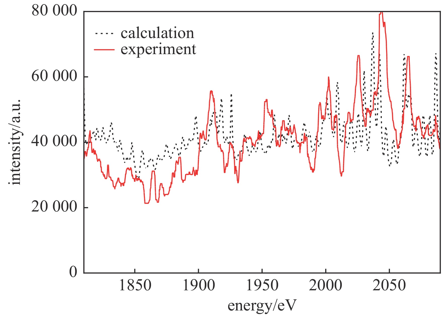
摘要: 时间分辨X射线吸收精细结构谱技术需要产生高亮度、均匀、宽光谱的X射线源。单一靶材产生的M带辐射源亮度高,但均匀性较差,因此提出了一种使用多种金属材料制备的多层膜复合靶产生M带辐射的方案。针对Si的K边X射线吸收谱实验,根据前期单一靶材M带光谱实验数据理论计算了最优的材料比例,制备了Au、Yb、Dy三种材料组成的多层膜复合靶,并在神光II激光装置上开展了脉冲激光驱动的多层膜复合靶辐射光谱测量,实验结果和理论计算基本一致。相比单一靶材,多层膜复合靶产生的M带辐射源具有光谱宽、整体亮度均匀的优点,在时间分辨X射线吸收精细结构谱中具有较大的应用潜力。Abstract: Time-resolved X-ray absorption fine structure spectrum technology needs to produce X-ray source with high brightness, uniform and wide spectrum. The M-band radiation source generated by an elementary target has high brightness, but poor uniformity. Therefore, this paper proposes a scheme to generate M-band radiation using a multilayer composite target prepared by a variety of metallic materials. For the K edge X-ray absorption spectroscopy experiment of Si, the optimal material ratio was theoretically calculated according to the previous elementary target M-band spectrum experiment data, and the multilayer composite target composed of Au, Yb, Dy was prepared. The radiation spectrum measurement of the multilayer composite target driven by pulse laser was carried out on the Shenguang II laser facility, and the experimental results were basically consistent with the theoretical calculation. Compared with an elementary target, the M-band radiation source generated by multilayer composite target has the advantages of wide spectrum and uniform overall brightness, and has great application potential in time-resolved X-ray absorption fine structure spectroscopy experiments.
-
Key words:
- spectroscopy /
- X-ray absorption fine structure /
- multilayer /
- composite target /
- M-band radiation
-
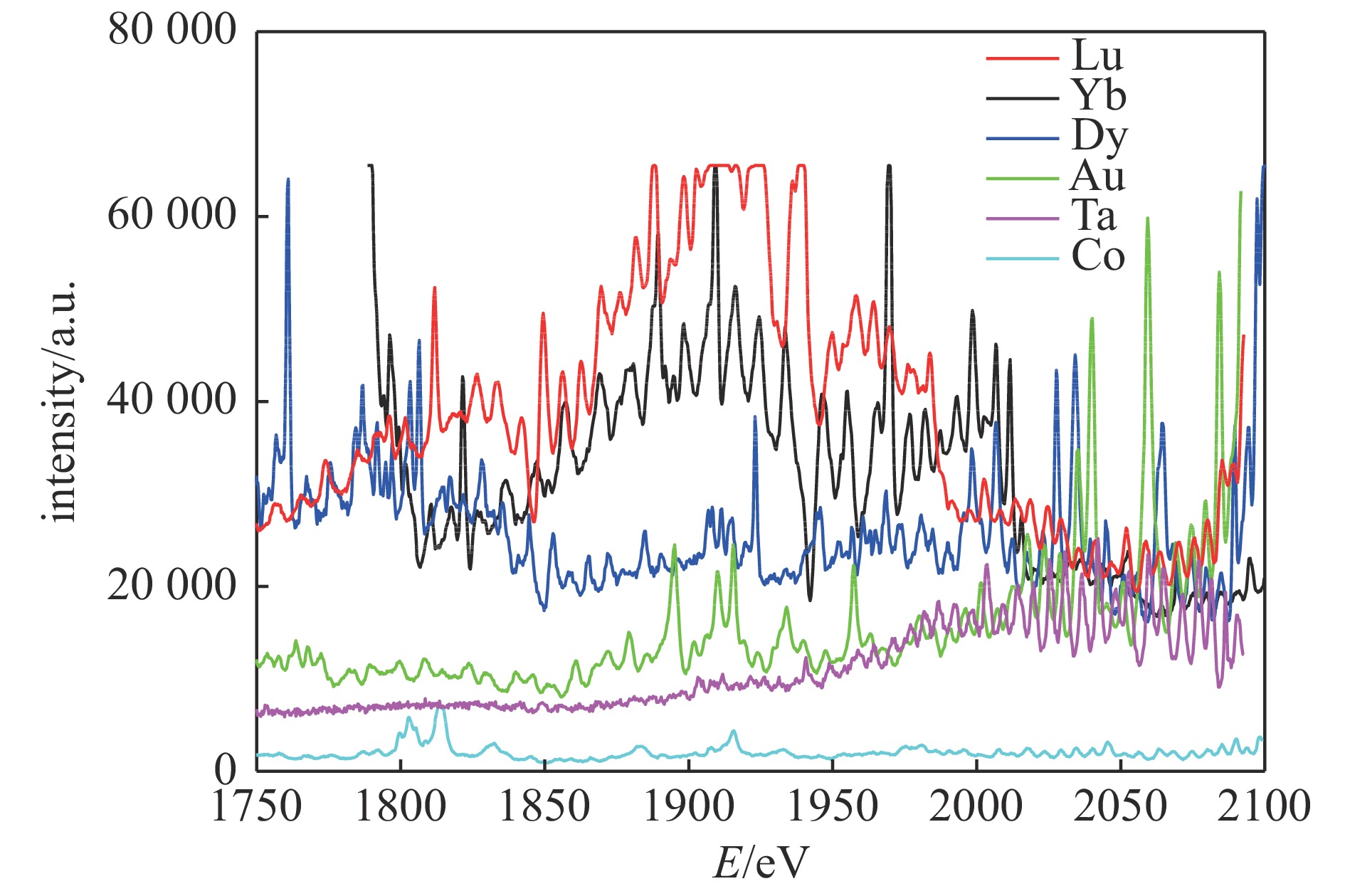
图 1 纳秒强激光辐照单一金属靶材产生的等离子体光谱
Figure 1. Plasma spectra generated by nanosecond high power laser irradiating on elementary metal target[16]
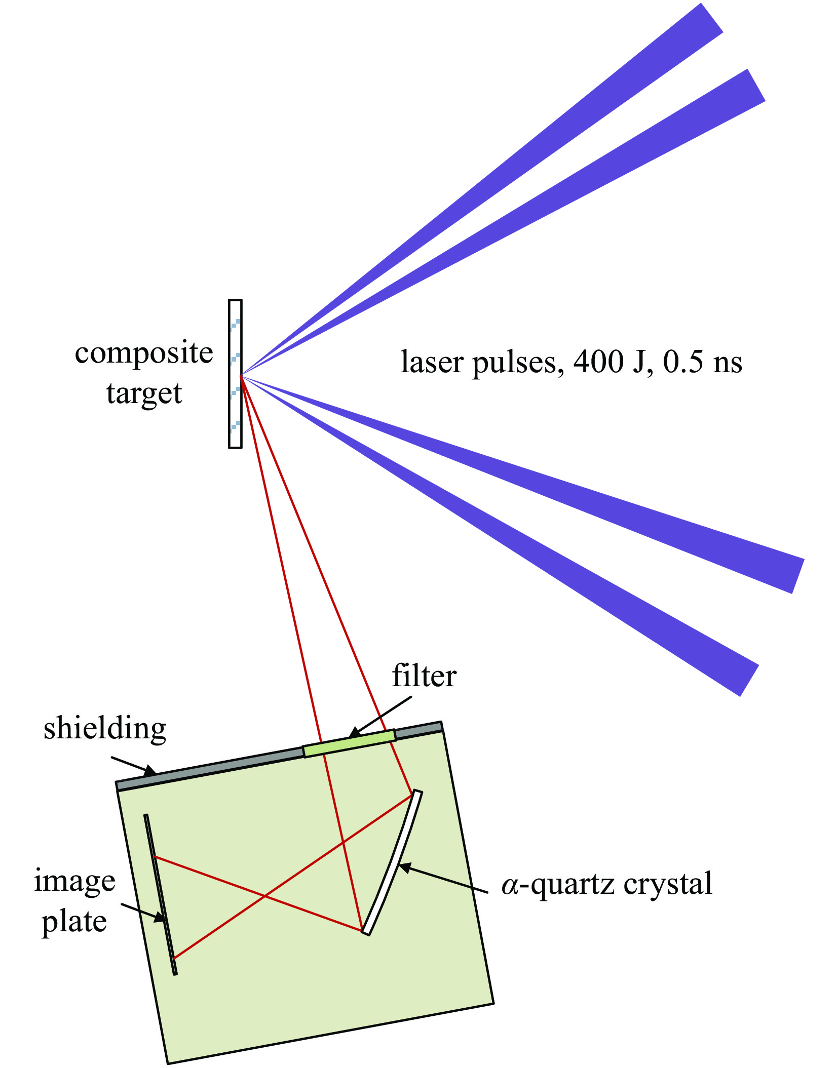
图 3 激光等离子体光谱测量实验布局示意图
Figure 3. Experimental layout of laser plasma spectrum measurement
-
[1] Bressler C, Chergui M. Ultrafast X-ray absorption spectroscopy[J]. Chemical Reviews, 2004, 104(4): 1781-812. doi: 10.1021/cr0206667 [2] Pertot Y, Schmidt C, Matthews M, et al. Time-resolved X-ray absorption spectroscopy with a water window high-harmonic source[J]. Science, 2017, 355(6322): 264. doi: 10.1126/science.aah6114 [3] Forget P, Dorchies F, Kieffer J C, et al. Ultrafast broadband laser plasma X-ray source for femtosecond time-resolved EXAFS[J]. Chemical Physics, 2004, 299(2-3): 259-263. doi: 10.1016/j.chemphys.2003.10.037 [4] Durr H A, Stamm C, Kachel T, et al. Ultrafast electron and spin dynamics in nickel probed with femtosecond X-ray pulses[J]. IEEE Transactions on Magnetics, 2008, 44(7): 1957-1961. doi: 10.1109/TMAG.2008.924544 [5] Pascarelli S, Mathon O, Mairs T, et al. The time-resolved and extreme-conditions XAS (TEXAS) facility at the european synchrotron radiation facility: the energy-dispersive x-ray absorption spectroscopy beamline ID24[J]. Journal of Synchrotron Radiation, 2016, 23(1): 353-368. doi: 10.1107/S160057751501783X [6] Pépin C. M, Torchio R, Occelli F, et al. White-line evolution in shocked solid Ta evidenced by synchrotron X-ray absorption spectroscopy[J]. Physical Review B, 2020, 102(14): 144102. doi: 10.1103/PhysRevB.102.144102 [7] Torchio R, Occelli F, Mathon O, et al. Probing local and electronic structure in warm dense matter: single pulse synchrotron X-ray absorption spectroscopy on shocked Fe[J]. Scientific Reports, 2016, 6: 26402. doi: 10.1038/srep26402 [8] Yaakobi B, Meyerhofer D D, Boehly T R, et al. Extended X-ray absorption fine structure measurements of laser shocks in Ti and V and phase transformation in Ti[J]. Physics of Plasmas, 2004, 11(5): 2688-2695. doi: 10.1063/1.1646673 [9] Yaakobi B, Boehly T R, Meyerhofer D D, et al. Extended X-ray absorption fine structure measurement of phase transformation in iron shocked by nanosecond laser[J]. Physics of Plasmas, 2005, 12(9): 1052. [10] Coppari F, Thorn D. B, Kemp G. E, et al. X-ray source development for EXAFS measurements on the National Ignition Facility.[J]. Review of Scientific Instruments, 2017, 88(8): 083907. doi: 10.1063/1.4999649 [11] Bolis R, Hernandez J-A, Recoules V, et al. X-ray absorption near edge spectroscopy study of warm dense MgO[J]. 2019, 26(11): 112703. [12] Benuzzi-Mounaix A, Dorchies F, Recoules V, et al. Electronic structure investigation of highly compressed aluminum with K edge absorption spectroscopy[J]. Physical Review Letters, 2011, 107(16): 165006. doi: 10.1103/PhysRevLett.107.165006 [13] Dorchies F, Lévy A, Goyon C, et al. Unraveling the solid-liquid-vapor phase transition dynamics at the atomic level with ultrafast X-ray absorption near-edge spectroscopy[J]. Physical Review Letters, 2011, 107(24): 245006. doi: 10.1103/PhysRevLett.107.245006 [14] Dorchies F, Fedorov N, Lecherbourg L. Experimental station for laser-based picosecond time-resolved X-ray absorption near-edge spectroscopy.[J]. Review of Scientific Instruments, 2015, 86(7): 073106. doi: 10.1063/1.4926348 [15] Zhao Y, Yang J, Zhang J, et al. K-shell photoabsorption edge of strongly coupled matter driven by laser-converted radiation.[J]. Physical Review Letters, 2013, 111(15): 155003. doi: 10.1103/PhysRevLett.111.155003 [16] 谭伯仲, 阳庆国, 刘冬兵, 等. 基于M壳层辐射的Si K边X射线吸收近边结构谱实验研究.[J]. 光学学报, 2018, 38(3):0330001 doi: 10.3788/AOS201838.0330001Tan B Z, Yang Q G, Liu D B, et al. Experimental study on Si K-edge X-ray absorption near-edge structure with M-shell radiation[J]. Acta Optica Sinica, 2018, 38(3): 0330001 doi: 10.3788/AOS201838.0330001 -

 下载:
下载:

 点击查看大图
图(4)
计量
- 文章访问数: 619
- HTML全文浏览量: 262
- PDF下载量: 50
- 被引次数: 0
