

Skin damage in mice induced by different power densities of 1064 nm laser
-
摘要: 通过实验和理论分析研究1064 nm激光不同输出功率对小鼠皮肤的热损伤规律。利用皮肤镜与光学相干断层成像(OCT)评估小鼠皮肤组织热损伤程度;参考Arrhenius热损伤方程进行理论分析,并与实验结果对比。结果显示,当激光连续辐照时间为400 ms时,激光输出功率密度小于958 W/cm2时,激光辐照处泛红;激光输出功率密度为958~1160 W/cm2时,损伤呈白色水疱状;激光输出功率密度为1160~1370 W/cm2时,损伤呈浅坑状焦黄斑,损伤斑周围伴一圈鼓起的白色皮肤水疱;激光的功率密度在1370~2190 W/cm2,损伤呈红色坑状斑,损伤斑周围伴黑黄色焦痂。
-
关键词:
- 1064 nm激光 /
- 皮肤热损伤 /
- Arrhenius热损伤方程 /
- 热损伤模型 /
- 热损伤分级
Abstract: The thermal damage of mouse skin under different output power of 1064 nm laser was studied by experiment and theoretical analysis. The degree of thermal injury of mouse skin tissue was evaluated by dermoscope and optical coherence tomography (OCT), and the theoretical analysis was made according to Arrhenius thermal damage equation, and the results were compared with the experimental results. The results show that when the laser’s continuous irradiation time is 400 ms and the laser output power density is less than 958 W/cm2, there is no obvious damage; when the laser output power density is 958−1160 W/cm2, the damage is white blister-like; when the laser output power density is 1160−1370 W/cm2, the damage is a shallow pit-like macula with a circle of bulging white skin blisters around the damage spot; when the laser power density is at 1370−2190 W/cm2, the damage is a red pit-like spot, and there is black-yellow eschar around the injury spot. -

图 1 激光辐照小鼠皮肤实验的示意图
Figure 1. Experimental diagram of laser irradiation on the skin of mouse

图 2 1064 nm激光辐照400 ms后小鼠皮肤创面的皮肤镜影像
Figure 2. Dermoscope images of injury in mice skin at different duration of 1064 nm laser irradiation (400 ms after irradiation; scale bar of dermoscope images: 1 mm)

图 3 不同功率密度激光辐照对小鼠皮肤损伤面积的影响
Figure 3. Effect of different power density laser irradiation on the skin lesion area of mice
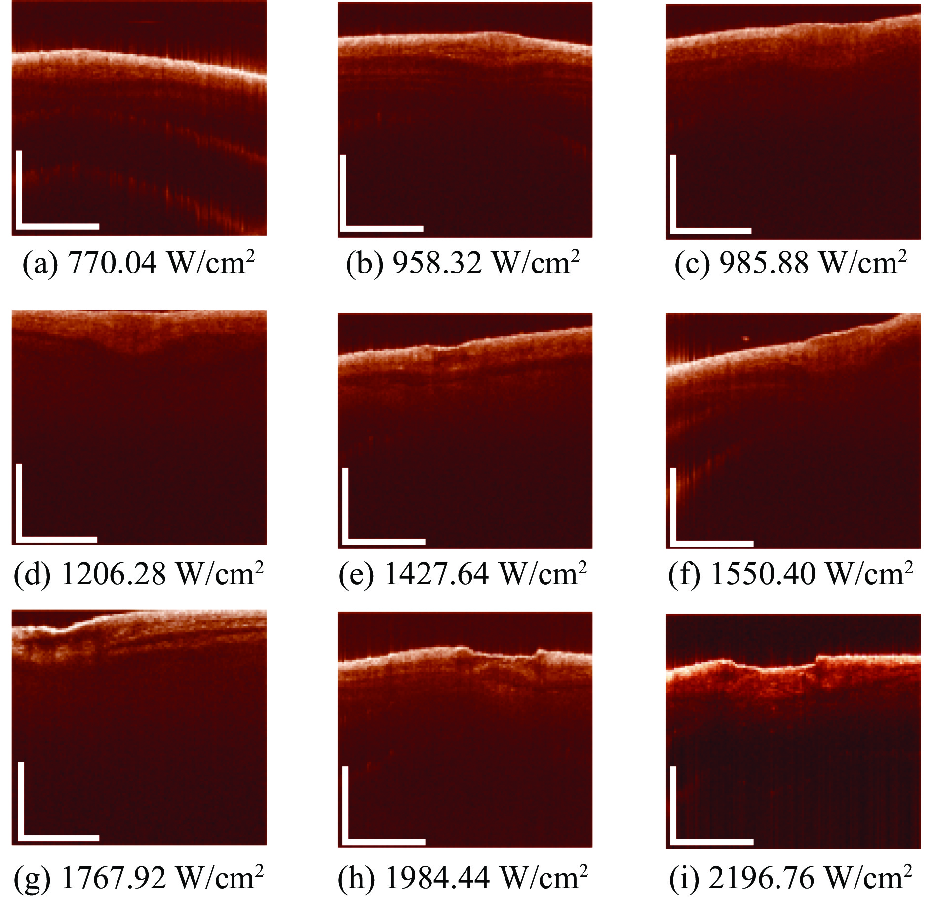
图 4 1064 nm激光辐照损伤后OCT结果观察(辐照400 ms,标尺:1 mm)
Figure 4. OCT images of injury in mice skin at different duration of 1064 nm laser irradiation(400 ms after irradiation; scale bar of OCT images:1 mm)
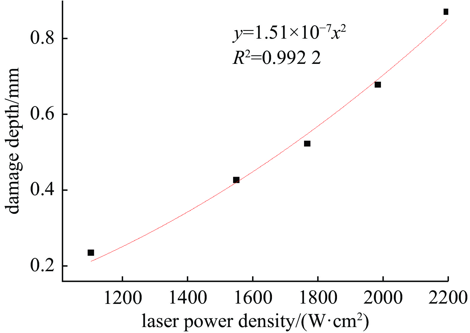
图 5 不同功率密度激光辐照对小鼠皮肤损伤深度的影响
Figure 5. Effect of laser irradiation with different power density on the depth of the skin damage in mice
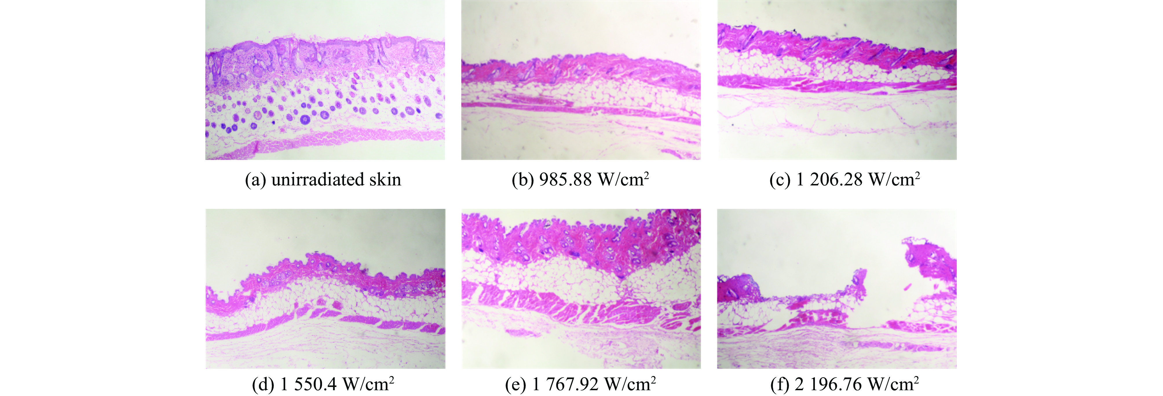
图 6 小鼠经1064 nm激光照射后皮肤创面的病理形态学变化(HE染色,×100)
Figure 6. Pathomorphological changes of skin wounds in mice irradiated by 1064nm laser (hematoxylin-eosin staining ×100)
表 1 1064 nm激光致生物皮肤组织皮肤损伤分级
Table 1. Grading of skin lesions in biological skin tissue by 1064 nm laser
laser irradiation power
density/(W·cm−2)damage appearance <958 firstdegree injury Redness at laser irradiation, recovered after some time. 958~1160 seconddegree injury White blisters, deformed and bulging (crumpled) skin around the injury. 1160~1370 thirddegree injury Vaporization of small amounts (or few lesions) on the superficial layer of the skin, blanching skin blisters with a bulging circle around the injury spot, and a shallow pit like focal macule. 1370~2190 fourthdegree injury Through the skin up to the fat layer, superficial vaporization was severe, splashing oil droplets, and around the injury with dark yellow eschar as red pit like spots.  下载: 导出CSV
表 2 皮肤热损伤程度与激光辐照功率对应关系
Table 2. The corresponding relationship between the degree of skin thermal injury and laser irradiation power
laser irradiation power density/(W·cm−2) Ω 374 1.80×10⁻⁵ 561 3.00×10⁻⁴ 762 1.9 958 14 1163 27 1369 111 1554 1200 1738 1.90×10⁸ 1934 5.50×10¹⁴
下载: 导出CSV
-
[1] 激光军事应用[J]. 中国光学与应用光学文摘, 2001, 15(5): 31-33Laser military application[J]. Chinese Optics, 2001, 15(5): 31-33 [2] 张延令. 激光在外科中的应用[J]. 国外医学. 外科学分册, 1984(3):143-146Zhang Yanling. Application of laser in surgery[J]. Foreign Medical Sciences. Surgery Section, 1984(3): 143-146 [3] 宋秋伦, 洪伟, 王倩. 逍遥丸联合调Q Nd: YAG 1064 nm激光治疗黄褐斑疗效分析[J]. 中国美容医学, 2022, 31(4):114-116Song Qiulun, Hong Wei, Wang Qian. Effect of Xiaoyao pill combined with Nd: YAG 1 064 nm laser on chloasma[J]. Chinese Journal of Aesthetic Medicine, 2022, 31(4): 114-116 [4] 左娜, 陶凯. 1064 nm Nd: YAG激光联合水光注射治疗面部皮肤老化疗效观察[J]. 中国美容医学, 2022, 31(5):23-25Zuo Na, Tao Kai. Efficacy of 1064 nm Nd: YAG laser combined with hyaluronic acid injection in the treatment of facial rejuvenation[J]. Chinese Journal of Aesthetic Medicine, 2022, 31(5): 23-25 [5] 施添霖. 积雪苷联合长脉宽1064 nm激光治疗瘢痕疙瘩的疗效观察[D]. 杭州: 浙江中医药大学, 2019Shi Tianlin. The therapeutic effect of asiaticoside ointment combined with long pulse 1064 nm laser on keloid[D]. Hangzhou: Zhejiang Chinese Medical University, 2019 [6] 冯微, 冯啸, 陈燕, 等. 长脉宽1064 nm激光在整形美容应用中的进展[J]. 中国美容整形外科杂志, 2021, 32(11):696-698 doi: 10.3969/j.issn.1673-7040.2021.11.016Feng Wei, Feng Xiao, Chen Yan, et al. Development of a 1064 nm long pulse width laser for cosmetic and cosmetic applications[J]. Chinese Journal of Aesthetic and Plastic Surgery, 2021, 32(11): 696-698 doi: 10.3969/j.issn.1673-7040.2021.11.016 [7] 李忠明, 骆清明. 激光对生物组织热和热致机械损伤的物理分析[J]. 激光生物学报, 2000, 9(2):81-84Li Zhongming, Luo Qingming. Physical analysis on tissues’ heat and heatinduced injury for laser irradiation[J]. Acta Laser Biology Sinica, 2000, 9(2): 81-84 [8] 王振华, 单健, 陈铀, 等. 激光对生物组织的热损伤机理研究及临床应用探讨[J]. 激光杂志, 2003, 24(1):60-62Wang Zhenhua, Shan Jian, Chen You, et al. A study on thermal damage of laser tissue and clinical application[J]. Laser Journal, 2003, 24(1): 60-62 [9] 吕晨阳, 战仁军. 激光-生物组织光热效应模型综述[J]. 激光杂志, 2021, 42(1):17-24Lv Chenyang, Zhan Renjun. Model review of laser-biological tissue photo-thermal effect[J]. Laser Journal, 2021, 42(1): 17-24 [10] 陈安宁, 吴家辉, 李希靖. 激光对生物组织热损伤的计算机模拟[J]. 光电子·激光, 2001, 12(2):212-214Chen Anning, Wu Jiahui, Li Xijing. Computer simulation of thermal damage to biological tissue during laser irradiation[J]. Journal of Optoelectronics·Laser, 2001, 12(2): 212-214 [11] 朱光明, 朱丹, 骆清铭, 等. 激光热疗中生物组织的光热特性响应以及动态热损伤研究[J]. 中国生物医学工程学报, 2004, 23(2):157-162Zhu Guangming, Zhu Dan, Luo Qingming, et al. Study on the optical-thermal response and dynamic thermal damage of bio-tissue during laser thermotherapy[J]. Chinese Journal of Biomedical Engineering, 2004, 23(2): 157-162 [12] 吴秀山, 侯宇, 李希靖. 强激光作用下生物组织热损伤问题的研究[J]. 中国计量学院学报, 2002, 13(2):135-138Wu Xiushan, Hou Yu, Li Xijing. The research on thermal damage in biological tissues induced by high-irradiance-laser[J]. Journal of China Jiliang University, 2002, 13(2): 135-138 [13] 周巡, 马琼, 刘智搏, 等. 1064 nm激光不同辐照时间对小鼠皮肤热损伤的实验与理论研究[J]. 强激光与粒子束, 2022, 34:011012 doi: 10.11884/HPLPB202234.210338Zhou Xun, Ma Qiong, Liu Zhibo, et al. 1064 nm laser induced thermal injure in mice skin with different laser duration[J]. High Power Laser and Particle Beams, 2022, 34: 011012 doi: 10.11884/HPLPB202234.210338 [14] Kagan R J, Peck M D, Ahrenholz D H, et al. Surgical management of the burn wound and use of skin substitutes: an expert panel white paper[J]. Journal of Burn Care & Research, 2013, 34(2): e60-e79. [15] Ganguly M, Mitra K. A modeling study to analyze thermal and mechanical effects of pulsed laser irradiation on tissues[J]. Computational Thermal Sciences: an International Journal, 2015, 7(5/6): 459-465. -


 点击查看大图
计量
- 文章访问数: 1640
- HTML全文浏览量: 521
- PDF下载量: 149
- 被引次数: 0
