| Citation: | Chu Wangsheng, Zhang Guobin, Sun Zhe, et al. Brief introduction of low-energy diffraction limited storage-ring-based synchrotron radiation and its applications[J]. High Power Laser and Particle Beams, 2022, 34: 104006. doi: 10.11884/HPLPB202234.220122

|

| [1] |
Brown G S, Moncton D E. Handbook on synchrotron radiation[M]. New York: North-Holland, 1991.
|
| [2] |
冼鼎昌. 神奇的光-同步辐射[M]. 长沙: 湖南教育出版社, 1994
Xian Dingchang. Synchrtron radiation—the magic light[M]. Changsha: Hunan Education Publishing House, 1994
|
| [3] |
Elder F R, Gurewitsch A M, Langmuir R V, et al. Radiation from electrons in a synchrotron[J]. Physical Review, 1947, 71(11): 829-830.
|
| [4] |
姜晓明, 修立松. 同步辐射及其应用[M]. 北京: 北京科学技术出版社, 1996
Jiang Xiaoming, Xiu Lisong. Synchrotron radiation and its applications[M]. Beijing: Beijing Science and Technology Press, 1996
|
| [5] |
马礼敦, 杨富家. 同步辐射应用概论[M]. 2版. 上海: 复旦大学出版社, 2005
Ma Lidun, Yang Fujia. Introduction to synchrotron radiation applications[M]. 2nd ed. Shanghai: Fudan University Press, 2005.
|
| [6] |
麦振洪. 同步辐射光的发展历史与现状——介绍新书《同步辐射光源及其应用》[J]. 现代物理知识, 2014, 26(2):65-71 doi: 10.13405/j.cnki.xdwz.2014.02.023
Mai Zhenhong. Development history and current situation of synchrotron radiation—introduction to the new book “synchrotron radiation source and its application”[J]. Modern Physics, 2014, 26(2): 65-71 doi: 10.13405/j.cnki.xdwz.2014.02.023
|
| [7] |
Hettel R. DLSR design and plans: an international overview[J]. Journal of Synchrotron Radiation, 2014, 21(5): 843-855. doi: 10.1107/S1600577514011515
|
| [8] |
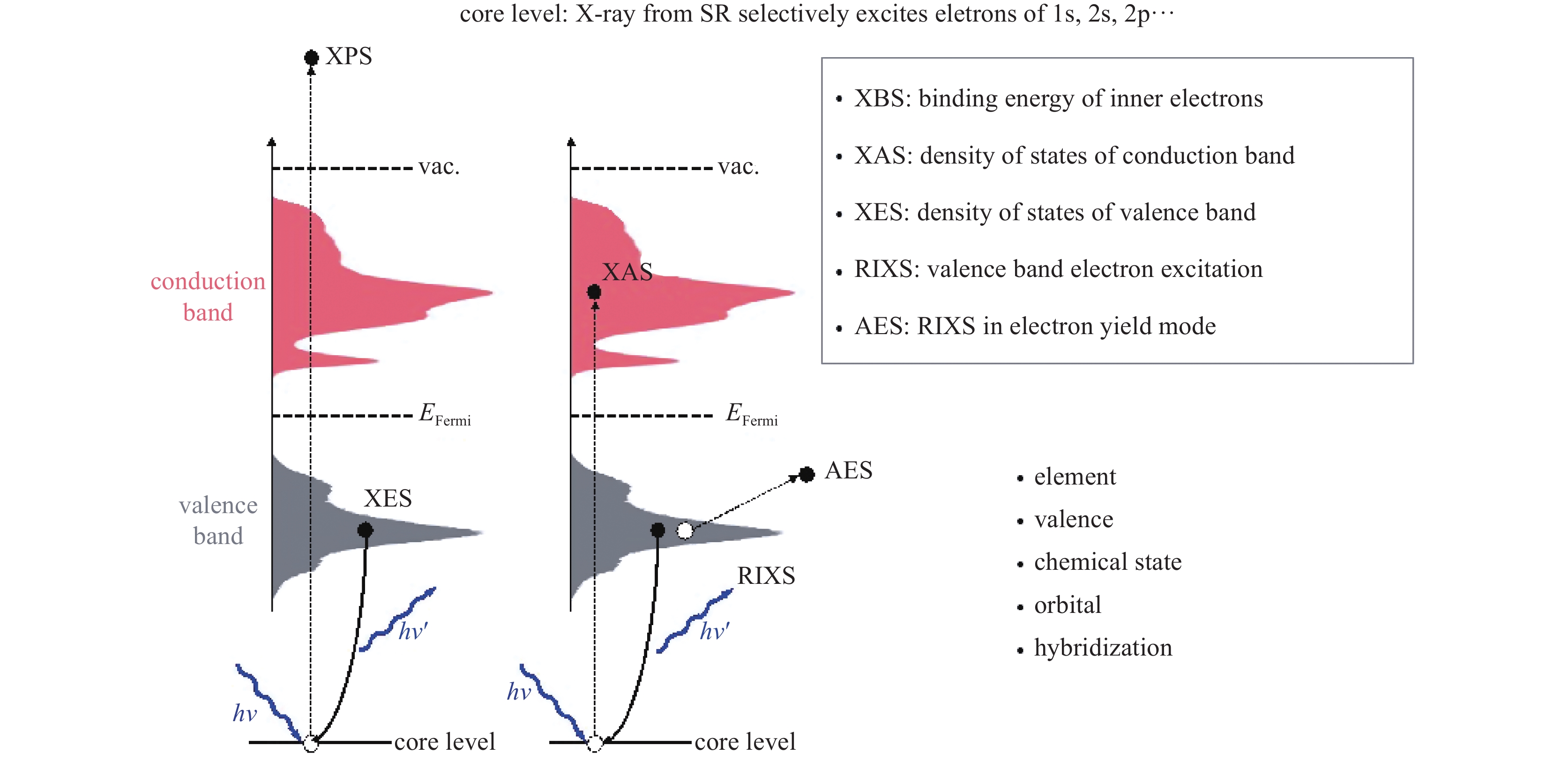
Hitchcock A P, Toney M F. Spectromicroscopy and coherent diffraction imaging: focus on energy materials applications[J]. Journal of Synchrotron Radiation, 2014, 21(5): 1019-1030. doi: 10.1107/S1600577514013046
|
| [9] |
Frenkel A I, van Bokhoven J A. X-ray spectroscopy for chemical and energy sciences: the case of heterogeneous catalysis[J]. Journal of Synchrotron Radiation, 2014, 21(5): 1084-1089. doi: 10.1107/S1600577514014854
|
| [10] |
de Jonge M D, Ryan C G, Jacobsen C J. X-ray nanoprobes and diffraction-limited storage rings: opportunities and challenges of fluorescence tomography of biological specimens[J]. Journal of Synchrotron Radiation, 2014, 21(5): 1031-1047. doi: 10.1107/S160057751401621X
|
| [11] |
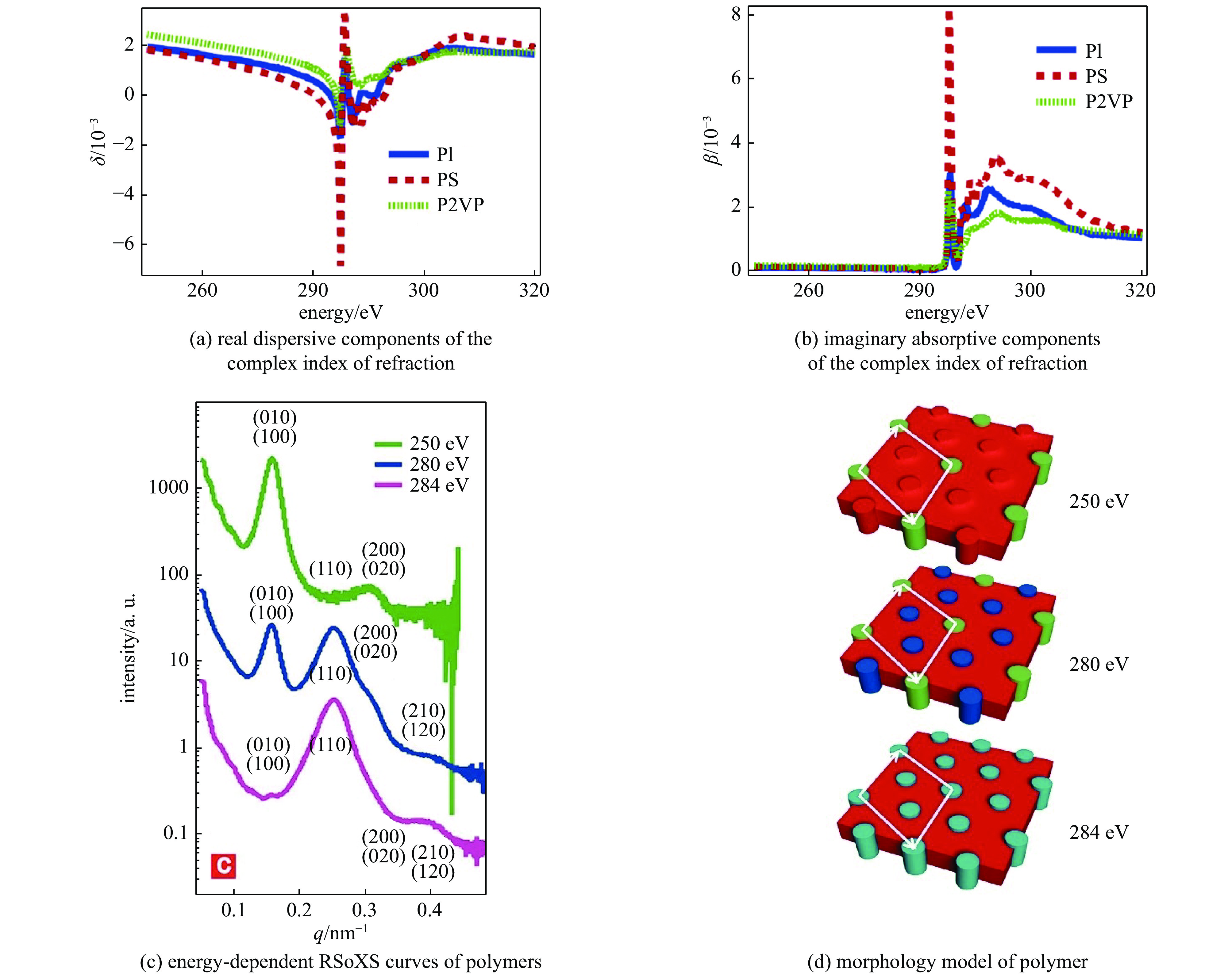
Liu Feng, Brady M A, Wang Cheng. Resonant soft X-ray scattering for polymer materials[J]. European Polymer Journal, 2016, 81: 555-568. doi: 10.1016/j.eurpolymj.2016.04.014
|
| [12] |
Comin R, Damascelli A. Resonant X-ray scattering studies of charge order in cuprates[J]. Annual Review of Condensed Matter Physics, 2016, 7: 369-405. doi: 10.1146/annurev-conmatphys-031115-011401
|
| [13] |
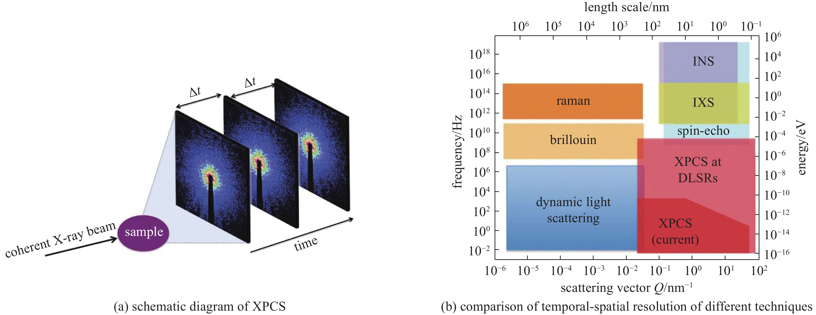
Shpyrko O G. X-ray photon correlation spectroscopy[J]. Journal of Synchrotron Radiation, 2014, 21(5): 1057-1064. doi: 10.1107/S1600577514018232
|
| [14] |
Sandy A R, Zhang Qingteng, Lurio L B. Hard X-ray photon correlation spectroscopy methods for materials studies[J]. Annual Review of Materials Research, 2018, 48: 167-190. doi: 10.1146/annurev-matsci-070317-124334
|
| [15] |
Tamarat P, Bodnarchuk M I, Trebbia J B, et al. The ground exciton state of formamidinium lead bromide perovskite nanocrystals is a singlet dark state[J]. Nature Materials, 2019, 18(7): 717-724. doi: 10.1038/s41563-019-0364-x
|
| [16] |
Ehrburger-Dolle F, Morfin I, Bley F, et al. XPCS investigation of the dynamics of filler particles in stretched filled elastomers[J]. Macromolecules, 2012, 45(21): 8691-8701. doi: 10.1021/ma3013674
|
| [17] |
Kukreja R, Hua N, Ruby J, et al. Orbital domain dynamics in magnetite below the Verwey transition[J]. Physical Review Letters, 2018, 121: 177601. doi: 10.1103/PhysRevLett.121.177601
|
| [18] |
Chen X M, Thampy V, Mazzoli C, et al. Remarkable stability of charge density wave order in La1.875Ba0.125CuO4[J]. Physical Review Letters, 2016, 117: 167001. doi: 10.1103/PhysRevLett.117.167001
|
| [19] |
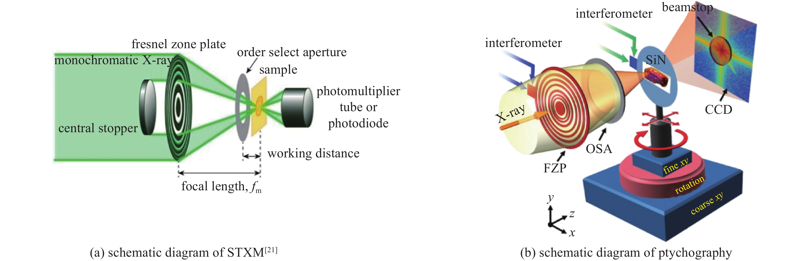
Kirz J, Rarback H. Soft X-ray microscopes[J]. Review of Scientific Instruments, 1985, 56(1): 1-13. doi: 10.1063/1.1138464
|
| [20] |
Pfeiffer F. X-ray ptychography[J]. Nature Photonics, 2018, 12(1): 9-17. doi: 10.1038/s41566-017-0072-5
|
| [21] |
Ohigashi T, Yuzawa H, Kosugi N. A low-pass filtering Fresnel zone plate for soft X-ray microscopic analysis down to the lithium K-edge region[J]. Review of Scientific Instruments, 2020, 91: 103110. doi: 10.1063/5.0020956
|
| [22] |
Chao Weilun, Harteneck B D, Liddle J A, et al. Soft X-ray microscopy at a spatial resolution better than 15 nm[J]. Nature, 2005, 435(7046): 1210-1213. doi: 10.1038/nature03719
|
| [23] |
Shapiro D A, Yu Y S, Tyliszczak T, et al. Chemical composition mapping with nanometre resolution by soft X-ray microscopy[J]. Nature Photonics, 2014, 8(10): 765-769. doi: 10.1038/nphoton.2014.207
|
| [24] |
Shi Xiaowen, Burdet N, Chen Bo, et al. X-ray ptychography on low-dimensional hard-condensed matter materials[J]. Applied Physical Reviews, 2019, 6: 011306. doi: 10.1063/1.5045131
|
| [25] |
Liu Xiaosong, Yang Wanli, Liu Zhi. Recent progress on synchrotron-based in-situ soft X-ray spectroscopy for energy materials[J]. Advanced Materials, 2014, 26(46): 7710-7729. doi: 10.1002/adma.201304676
|
| [26] |
Berkeley Lab, U. S. Department of Energy Office of Science, ALS-U. ALS-U: solving scientific challenges with coherent soft X-rays, workshop report on early science enabled by the Advanced Light Source Upgrade[R]. 2017.
|
| [27] |
Avila J, Asensio M C. First NanoARPES user facility available at SOLEIL: an innovative and powerful tool for studying advanced materials[J]. Synchrotron Radiation News, 2014, 27(2): 24-30. doi: 10.1080/08940886.2014.889549
|
| [28] |
Kastl C, Koch R J, Chen C T, et al. Effects of defects on band structure and excitons in WS2 revealed by nanoscale photoemission spectroscopy[J]. ACS Nano, 2019, 13(2): 1284-1291.
|
| [29] |
Jia Chunjing, Wohlfeld K, Wang Yao, et al. Using RIXS to uncover elementary charge and spin excitations[J]. Physical Review X, 2016, 6: 021020.
|
| [30] |
Qiao Ruimin, Li Qinghao, Zhuo Zengqing, et al. High-efficiency in situ resonant inelastic X-ray scattering (iRIXS) endstation at the Advanced Light Source[J]. Review of Scientific Instruments, 2017, 88: 033106. doi: 10.1063/1.4977592
|
| [31] |
Chuang Yide, Feng Xuefei, Glans-Suzuki P A, et al. A design of resonant inelastic X-ray scattering (RIXS) spectrometer for spatial- and time-resolved spectroscopy[J]. Journal of Synchrotron Radiation, 2020, 27(3): 695-707. doi: 10.1107/S1600577520004440
|
| [32] |
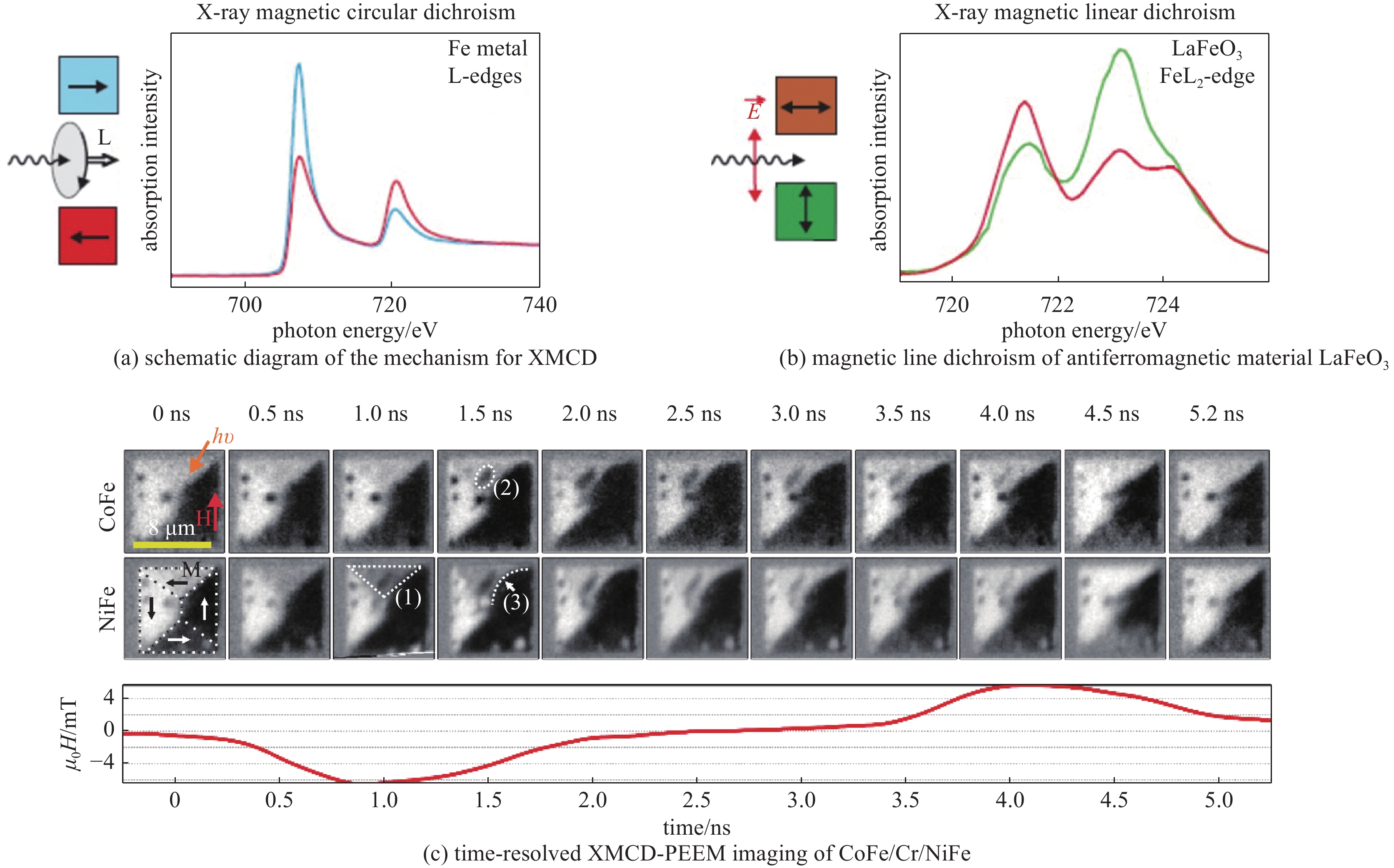
Kaiser A M, Schöppner C, Römer F M, et al. Nano and picosecond magnetization dynamics of weakly coupled CoFe/Cr/NiFe trilayers studied by a multitechnique approach[J]. Physical Review B, 2011, 84: 134406. doi: 10.1103/PhysRevB.84.134406
|
| [33] |
van der Laan G, Figueroa A I. X-ray magnetic circular dichroism—A versatile tool to study magnetism[J]. Coordination Chemistry Reviews, 2014, 277/278: 95-129. doi: 10.1016/j.ccr.2014.03.018
|
| [34] |
吴义政. 同步辐射X射线磁二色性在自旋电子学研究中的应用[J]. 物理, 2010, 39(6):406-415
Wu Yizheng. Applications of X-ray magnetic dichroism in spintronics[J]. Wuli, 2010, 39(6): 406-415
|
| [35] |
Mengotti E, Heyderman L J, Rodríguez A F, et al. Real-space observation of emergent magnetic monopoles and associated Dirac strings in artificial kagome spin ice[J]. Nature Physics, 2011, 7(1): 68-74. doi: 10.1038/nphys1794
|
| [36] |
Zhao Tong, Scholl A, Zavaliche F, et al. Electrical control of antiferromagnetic domains in multiferroic BiFeO3 films at room temperature[J]. Nature Materials, 2006, 5(10): 823-829. doi: 10.1038/nmat1731
|
| [37] |
Suchorski Y, Kozlov S M, Bespalov I, et al. The role of metal/oxide interfaces for long-range metal particle activation during CO oxidation[J]. Nature Materials, 2018, 17(6): 519-522. doi: 10.1038/s41563-018-0080-y
|
| [38] |
Shiino T, Oh S H, Haney P M, et al. Antiferromagnetic domain wall motion driven by spin-orbit torques[J]. Physical Review Letters, 2016, 117: 087203. doi: 10.1103/PhysRevLett.117.087203
|
| [39] |
Litzius K, Lemesh I, Krüger B, et al. Skyrmion Hall effect revealed by direct time-resolved X-ray microscopy[J]. Nature Physics, 2017, 13(2): 170-175. doi: 10.1038/nphys4000
|
| [40] |
Wang Wenbo, Ou Yunbo, Liu Chang, et al. Direct evidence of ferromagnetism in a quantum anomalous Hall system[J]. Nature Physics, 2018, 14(8): 791-795. doi: 10.1038/s41567-018-0149-1
|
| [41] |
Jiang Wanjun, Upadhyaya P, Zhang Wei, et al. Blowing magnetic skyrmion bubbles[J]. Science, 2015, 349(6245): 283-286. doi: 10.1126/science.aaa1442
|
| [42] |
Fukami S, Zhang Chaoliang, DuttaGupta S, et al. Magnetization switching by spin-orbit torque in an antiferromagnet-ferromagnet bilayer system[J]. Nature Materials, 2016, 15(5): 535-541. doi: 10.1038/nmat4566
|
| [43] |
Zhou Jiadong, Lin Junhao, Huang Xiangwei, et al. A library of atomically thin metal chalcogenides[J]. Nature, 2018, 556(7701): 355-359. doi: 10.1038/s41586-018-0008-3
|
| [44] |
Baltz V, Manchon A, Tsoi M, et al. Antiferromagnetic spintronics[J]. Reviews of Modern Physics, 2018, 90: 015005. doi: 10.1103/RevModPhys.90.015005
|
| [45] |
Lesne E, Fu Yu, Oyarzun S, et al. Highly efficient and tunable spin-to-charge conversion through Rashba coupling at oxide interfaces[J]. Nature Materials, 2016, 15(12): 1261-1266. doi: 10.1038/nmat4726
|
| [46] |
Dagotto E. Complexity in strongly correlated electronic systems[J]. Science, 2005, 309(5732): 257-262. doi: 10.1126/science.1107559
|
| [47] |
Keimer B, Moore J E. The physics of quantum materials[J]. Nature Physics, 2017, 13(11): 1045-1055. doi: 10.1038/nphys4302
|
| [48] |
Tokura Y, Kawasaki M, Nagaosa N. Emergent functions of quantum materials[J]. Nature Physics, 2017, 13(11): 1056-1068. doi: 10.1038/nphys4274
|
| [49] |
Basov D N, Averitt R D, Hsieh D. Towards properties on demand in quantum materials[J]. Nature Materials, 2017, 16(11): 1077-1088. doi: 10.1038/nmat5017
|
| [50] |
Borisenko S V, Evtushinsky D V, Liu Zhonghao, et al. Direct observation of spin–orbit coupling in iron-based superconductors[J]. Nature Physics, 2016, 12(4): 311-317. doi: 10.1038/nphys3594
|
| [51] |
Lu Donghui, Vishik IM, Yi Ming, et al. Angle-resolved photoemission studies of quantum materials[J]. Annual Review of Condensed Matter Physics, 2012, 3: 129-167. doi: 10.1146/annurev-conmatphys-020911-125027
|
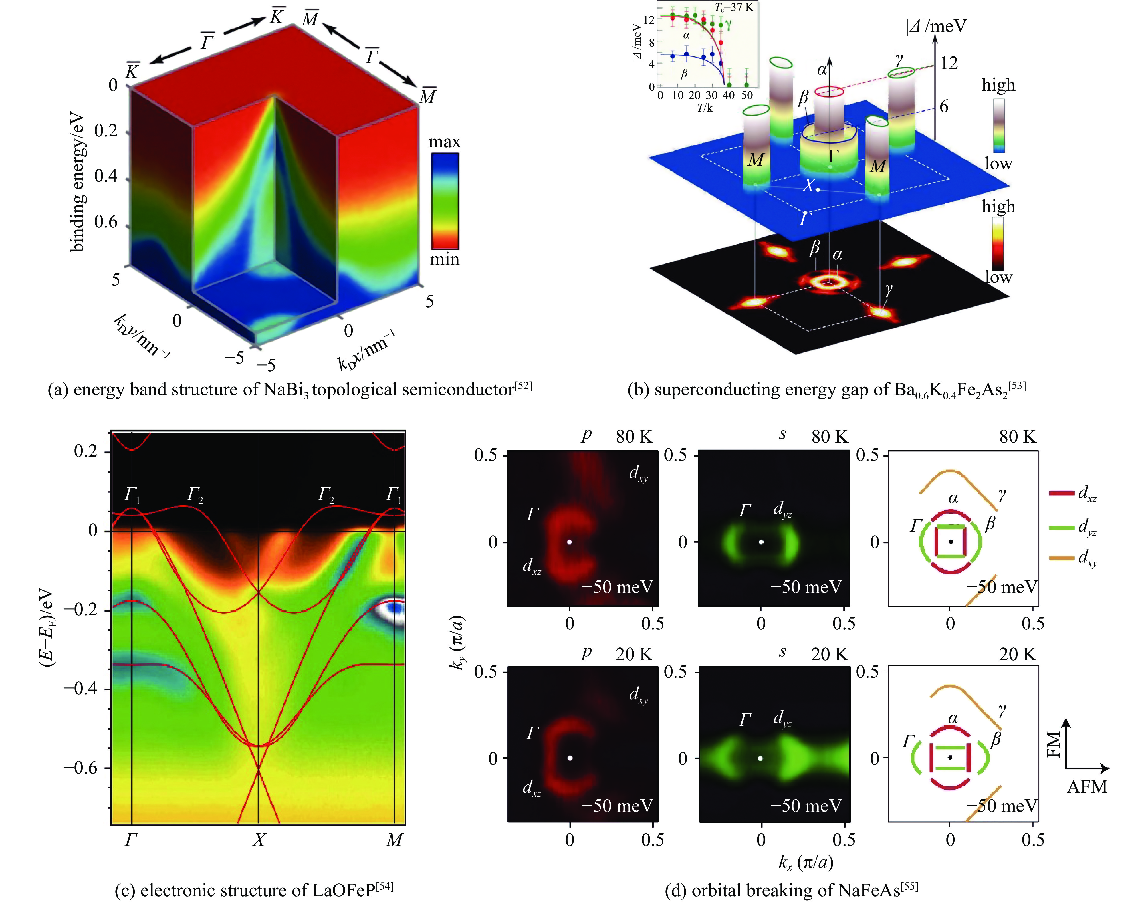
| [52] |
Liu Z K, Zhou B, Zhang Yi, et al. Discovery of a three-dimensional topological dirac semimetal, Na3Bi[J]. Science, 2014, 343(6173): 864-867. doi: 10.1126/science.1245085
|
| [53] |
Ding Hanjie, Richard P, Nakayama K, et al. Observation of Fermi-surface–dependent nodeless superconducting gaps in Ba0.6K0.4Fe2As2[J]. Europhysics Letters, 2008, 83: 47001. doi: 10.1209/0295-5075/83/47001
|
| [54] |
Lu D H, Yi Ming, Mo S K, et al. Electronic structure of the iron-based superconductor LaOFeP[J]. Nature, 2008, 455(7209): 81-84. doi: 10.1038/nature07263
|
| [55] |
Zhang Yong, He Cheng, Ye Z R, et al. Symmetry breaking via orbital-dependent reconstruction of electronic structure in detwinned NaFeAs[J]. Physical Review B, 2012, 85: 085121. doi: 10.1103/PhysRevB.85.085121
|
| [56] |
Zhou Shuyun, Gweon GH, Fedorov AV, et al. Erratum: substrate-induced bandgap opening in epitaxial graphene[J]. Nature Materials, 2007, 6: 916. doi: 10.1038/nmat2056
|
| [57] |
Fäth M, Freisem S, Menovsky A A, et al. Spatially inhomogeneous metal-insulator transition in doped manganites[J]. Science, 1999, 285(5433): 1540-1542. doi: 10.1126/science.285.5433.1540
|
| [58] |
Song Canli, Wang Yilin, Cheng Peng, et al. Direct observation of nodes and twofold symmetry in FeSe superconductor[J]. Science, 2011, 332(6036): 1410-1413. doi: 10.1126/science.1202226
|
| [59] |
Li Wei, Ding Hao, Deng Peng, et al. Phase separation and magnetic order in K-doped iron selenide superconductor[J]. Nature Physics, 2012, 8(2): 126-130. doi: 10.1038/nphys2155
|
| [60] |
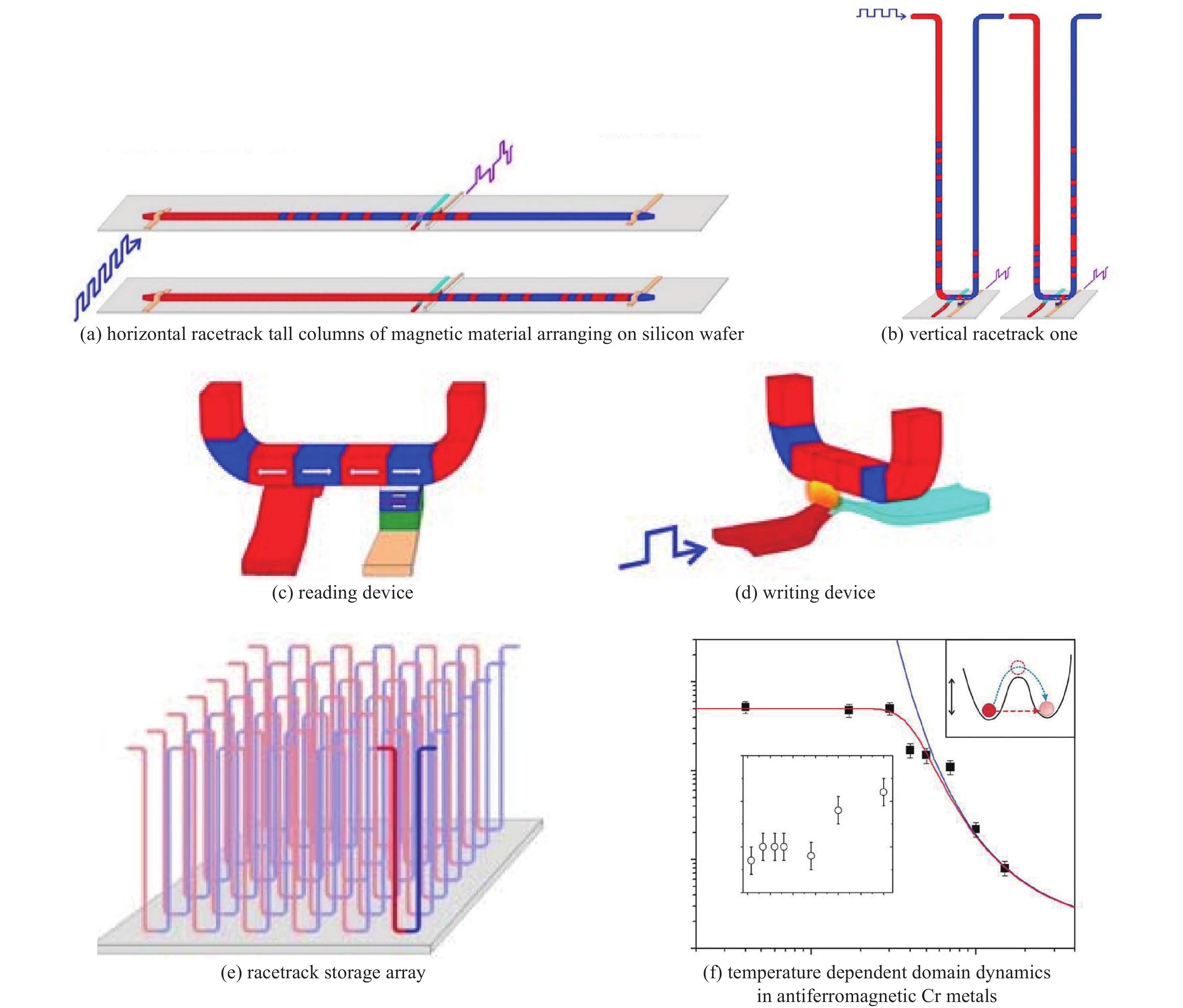
Parkin S. Racetrack memory: a storage class memory based on current controlled magnetic domain wall motion[C]//2009 Device Research Conference. 2009: 3-6.
|
| [61] |
Knafo W, Raymond S, Lejay P, et al. Antiferromagnetic criticality at a heavy-fermion quantum phase transition[J]. Nature Physics, 2009, 5(10): 753-757. doi: 10.1038/nphys1374
|
| [62] |
Schröder A, Aeppli G, Coldea R, et al. Onset of antiferromagnetism in heavy-fermion metals[J]. Nature, 2000, 407(6802): 351-355. doi: 10.1038/35030039
|
| [63] |
Duan Chunruo, Baumbach R E, Podlesnyak A, et al. Resonance from antiferromagnetic spin fluctuations for superconductivity in UTe2[J]. Nature, 2021, 600(7890): 636-640. doi: 10.1038/s41586-021-04151-5
|
| [64] |
Shpyrko O G, Isaacs E D, Logan J M, et al. Direct measurement of antiferromagnetic domain fluctuations[J]. Nature, 2007, 447(7140): 68-71. doi: 10.1038/nature05776
|
| [65] |
Kim K J, Kim S K, Hirata Y, et al. Fast domain wall motion in the vicinity of the angular momentum compensation temperature of ferrimagnets[J]. Nature Materials, 2017, 16(12): 1187-1192. doi: 10.1038/nmat4990
|
| [66] |
Bernstein D P, Bräuer B, Kukreja R, et al. Nonuniform switching of the perpendicular magnetization in a spin-torque-driven magnetic nanopillar[J]. Physical Review B, 2011, 83: 180410. doi: 10.1103/PhysRevB.83.180410
|
| [67] |
Reyren N, Bouzehouane K, Chauleau J Y, et al. Skyrmions in magnetic multilayers: chirality, electrical detection and current-induced motion[C]. Proceedings of SPIE, Spintronics X. 2017: 1035724.
|
| [68] |
Hellman F, Hoffmann A, Tserkovnyak Y, et al. Interface-induced phenomena in magnetism[J]. Reviews of Modern Physics, 2017, 89: 025006. doi: 10.1103/RevModPhys.89.025006
|
| [69] |
Banerjee S, Erten O, Randeria M. Ferromagnetic exchange, spin-orbit coupling and spiral magnetism at the LaAlO3/SrTiO3 interface[J]. Nature Physics, 2013, 9(10): 626-630. doi: 10.1038/nphys2702
|
| [70] |
Grisolia M N, Varignon J, Sanchez-Santolino G, et al. Hybridization-controlled charge transfer and induced magnetism at correlated oxide interfaces[J]. Nature Physics, 2016, 12(5): 484-492. doi: 10.1038/nphys3627
|
| [71] |
Saito Y, Nakamura Y, Bahramy M S, et al. Superconductivity protected by spin-valley locking in ion-gated MoS2[J]. Nature Physics, 2016, 12(2): 144-149. doi: 10.1038/nphys3580
|
| [72] |
Li J, Shelford L R, Shafer P, et al. Direct detection of pure ac spin current by X-ray pump-probe measurements[J]. Physical Review Letters, 2016, 117: 076602. doi: 10.1103/PhysRevLett.117.076602
|
| [73] |
Li Wenjing, Bykova I, Zhang Shilei, et al. Anatomy of skyrmionic textures in magnetic multilayers[J]. Advanced Materials, 2019, 31: 1807683. doi: 10.1002/adma.201807683
|
| [74] |
Tumbleston J R, Collins B A, Yang Liqiang, et al. The influence of molecular orientation on organic bulk heterojunction solar cells[J]. Nature Photonics, 2014, 8(5): 385-391. doi: 10.1038/nphoton.2014.55
|
| [75] |
Collins B A, Cochran J E, Yan Hongping, et al. Polarized X-ray scattering reveals non-crystalline orientational ordering in organic films[J]. Nature Materials, 2012, 11(6): 536-543. doi: 10.1038/nmat3310
|
| [76] |
Suh H S, Kang Huiman, Nealey P F, et al. Thickness dependence of neutral parameter windows for perpendicularly oriented block copolymer thin films[J]. Macromolecules, 2010, 43(10): 4744-4751. doi: 10.1021/ma100150j
|
| [77] |
Ahn H, Shin C, Lee B, et al. Phase transitions of block copolymer film on homopolymer-grafted substrate[J]. Macromolecules, 2010, 43(4): 1958-1963. doi: 10.1021/ma9022229
|
| [78] |
Sivaniah E, Hayashi Y, Matsubara S, et al. Symmetric diblock copolymer thin films on rough substrates. Kinetics and structure formation in pure block copolymer thin films[J]. Macromolecules, 2005, 38(5): 1837-1849. doi: 10.1021/ma0482157
|
| [79] |
Hur S M, Khaira G S, Ramírez-Hernández A, et al. Simulation of defect reduction in block copolymer thin films by solvent annealing[J]. ACS Macro Letters, 2015, 4(1): 11-15. doi: 10.1021/mz500705q
|
| [80] |
Sinturel C, Vayer M, Morris M, et al. Solvent vapor annealing of block polymer thin films[J]. Macromolecules, 2013, 46(14): 5399-5415. doi: 10.1021/ma400735a
|
| [81] |
De Rosa C, Park C, Thomas E L, et al. Microdomain patterns from directional eutectic solidification and epitaxy[J]. Nature, 2000, 405(6785): 433-437. doi: 10.1038/35013018
|
| [82] |
Tang Chuanbing, Wu Wei, Smilgies D M, et al. Robust control of microdomain orientation in thin films of block copolymers by zone casting[J]. Journal of the American Chemical Society, 2011, 133(30): 11802-11809. doi: 10.1021/ja204724h
|
| [83] |
Saito I, Miyazaki T, Yamamoto K. Depth-resolved structure analysis of cylindrical microdomain in block copolymer thin film by grazing-incidence small-angle X-ray scattering utilizing low-energy x-rays[J]. Macromolecules, 2015, 48(22): 8190-8196. doi: 10.1021/acs.macromol.5b01883
|
| [84] |
Gann E, Watson A, Tumbleston J R, et al. Topographic measurement of buried thin-film interfaces using a grazing resonant soft X-ray scattering technique[J]. Physical Review B, 2014, 90: 245421. doi: 10.1103/PhysRevB.90.245421
|
| [85] |
Leheny R L. XPCS: nanoscale motion and rheology[J]. Current Opinion in Colloid & Interface Science, 2012, 17(1): 3-12.
|
| [86] |
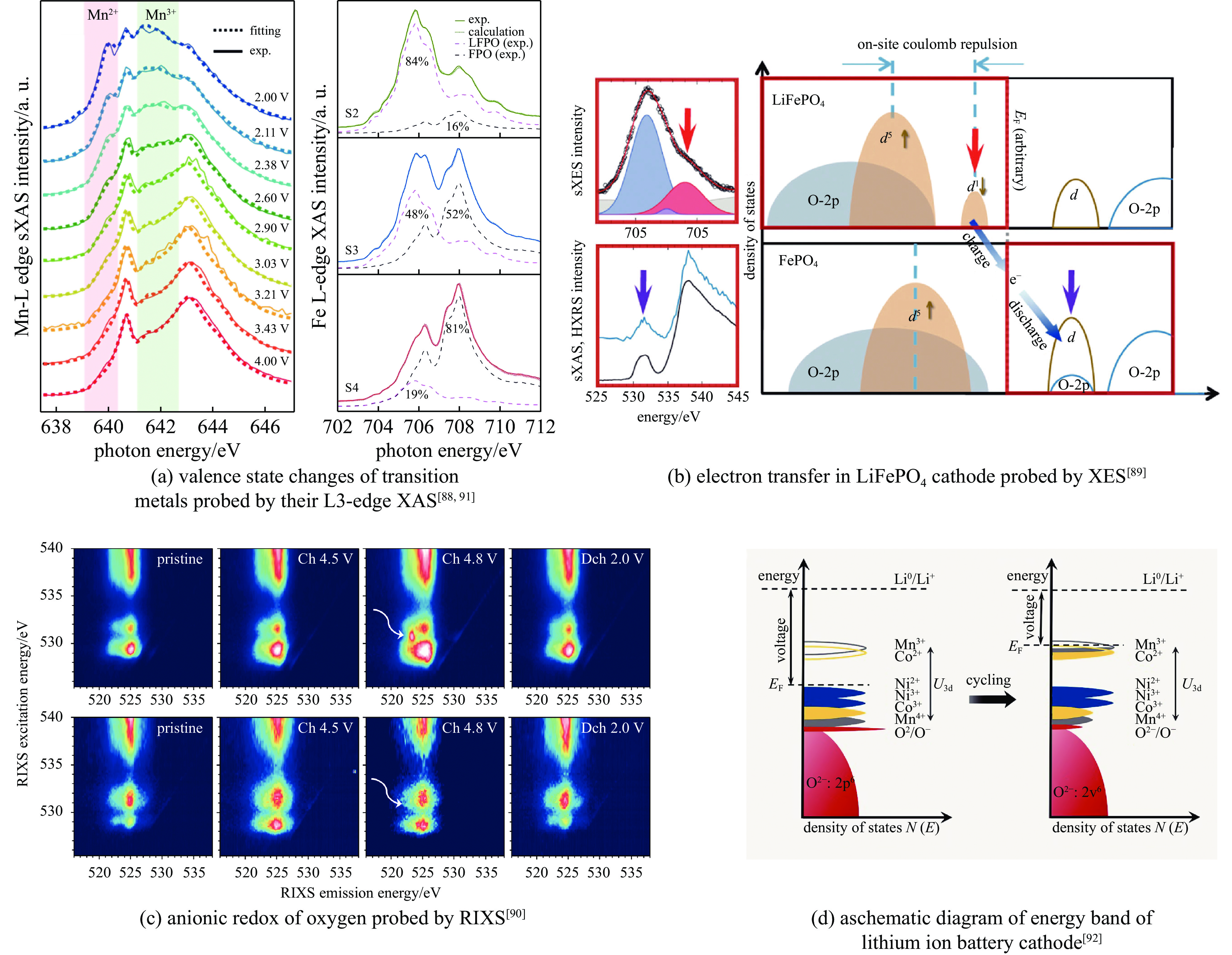
Lu Jun, Wu Tianpin, Amine K. State-of-the-art characterization techniques for advanced lithium-ion batteries[J]. Nature Energy, 2017, 2: 17011. doi: 10.1038/nenergy.2017.11
|
| [87] |
Peled E, Menkin S. Review—SEI: past, present and future[J]. Journal of the Electrochemical Society, 2017, 164(7): A1703-A1719. doi: 10.1149/2.1441707jes
|
| [88] |
Liu Xiaosong, Liu Jun, Qiao Ruimin, et al. Phase transformation and lithiation effect on electronic structure of LixFePO4: an in-depth study by soft X-ray and simulations[J]. Journal of the American Chemical Society, 2012, 134(33): 13708-13715. doi: 10.1021/ja303225e
|
| [89] |
Liu Xiaosong, Wang Y J, Barbiellini B, et al. Why LiFePO4 is a safe battery electrode: Coulomb repulsion induced electron-state reshuffling upon lithiation[J]. Physical Chemistry Chemical Physics, 2015, 17(39): 26369-26377. doi: 10.1039/C5CP04739K
|
| [90] |
Xu Jing, Sun Meiling, Qiao Ruimin, et al. Elucidating anionic oxygen activity in lithium-rich layered oxides[J]. Nature Communications, 2018, 9: 947. doi: 10.1038/s41467-018-03403-9
|
| [91] |
Wu Jinpeng, Shen Zhixun, Yang Wanli. Redox mechanism in Na-ion battery cathodes probed by advanced soft X-ray spectroscopy[J]. Frontiers in Chemistry, 2020, 8: 816. doi: 10.3389/fchem.2020.00816
|
| [92] |
Zhao Shuoqing, Yan Kang, Zhang Jinqiang, et al. Reaction mechanisms of layered lithium-rich cathode materials for high-energy lithium-ion batteries[J]. Angewandte Chemie International Edition, 2021, 60(5): 2208-2220. doi: 10.1002/anie.202000262
|
| [93] |
House R A, Maitra U, Pérez-Osorio M A, et al. Superstructure control of first-cycle voltage hysteresis in oxygen-redox cathodes[J]. Nature, 2020, 577(7791): 502-508. doi: 10.1038/s41586-019-1854-3
|
| [94] |
Yang Chunpeng, Fu Kun, Zhang Ying, et al. Protected lithium-metal anodes in batteries: from liquid to solid[J]. Advanced Materials, 2017, 29: 1701169. doi: 10.1002/adma.201701169
|
| [95] |
Li Yiyang, Weker J N, Gent W E, et al. Dichotomy in the lithiation pathway of ellipsoidal and platelet LiFePO4 particles revealed through nanoscale operando state-of-charge imaging[J]. Advanced Functional Materials, 2015, 25(24): 3677-3687. doi: 10.1002/adfm.201500286
|
| [96] |
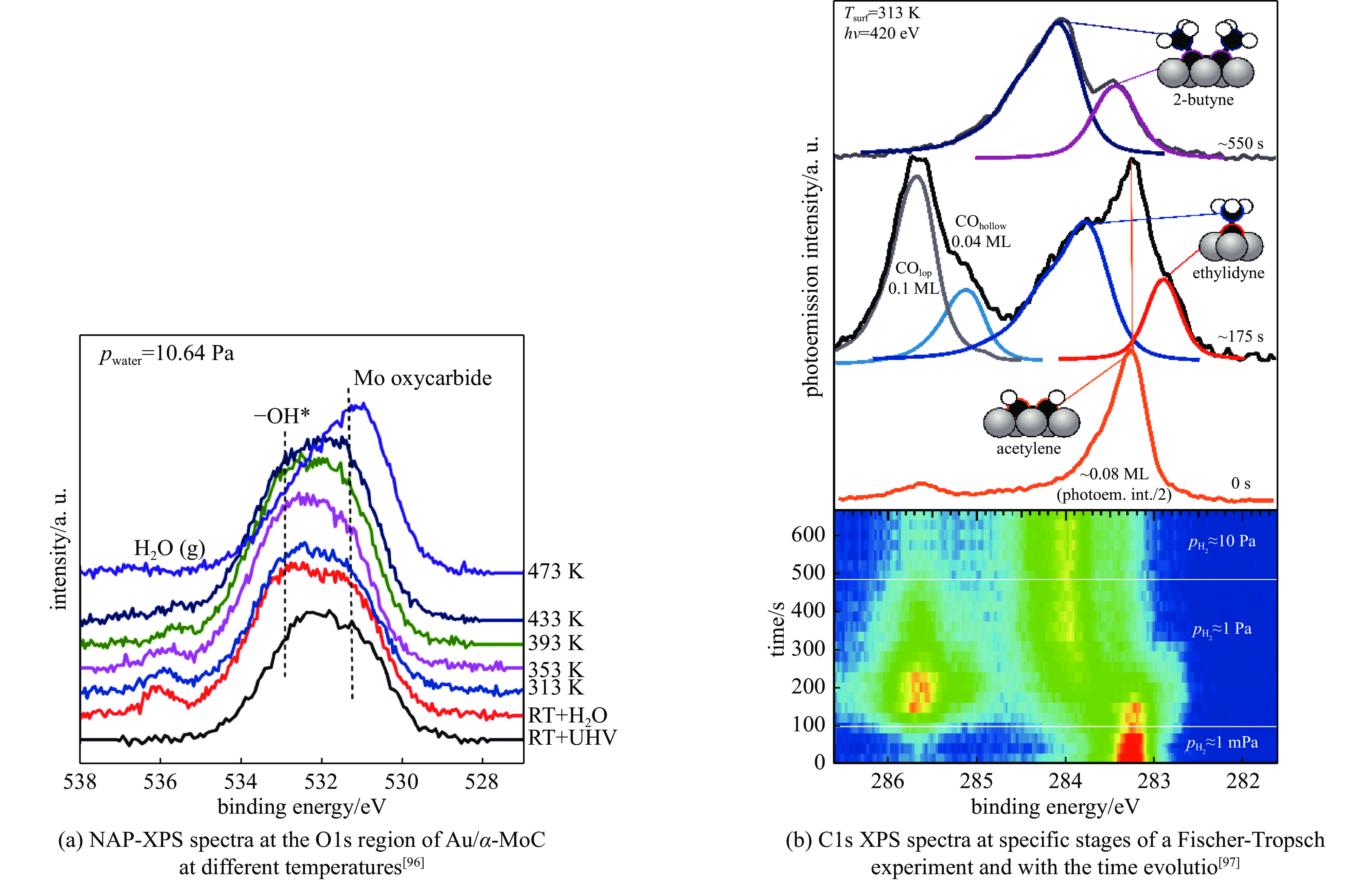
Yao Siyu, Zhang Xiao, Zhou Wu, et al. Atomic-layered Au clusters on α-MoC as catalysts for the low-temperature water-gas shift reaction[J]. Science, 2017, 357(6349): 389-393. doi: 10.1126/science.aah4321
|
| [97] |
Weststrate C J, Sharma D, Rodriguez D G, et al. Mechanistic insight into carbon-carbon bond formation on cobalt under simulated Fischer-Tropsch synthesis conditions[J]. Nature Communications, 2020, 11: 750. doi: 10.1038/s41467-020-14613-5
|
| [98] |
Zou Xiaoxin, Zhang Yu. Noble metal-free hydrogen evolution catalysts for water splitting[J]. Chemical Society Reviews, 2015, 44(15): 5148-5180. doi: 10.1039/C4CS00448E
|
| [99] |
Favaro M, Yang Jinhui, Nappini S, et al. Understanding the oxygen evolution reaction mechanism on CoOx using Operando ambient-pressure X-ray photoelectron spectroscopy[J]. Journal of the American Chemical Society, 2017, 139(26): 8960-8970. doi: 10.1021/jacs.7b03211
|
| [100] |
Su Xiaozhi, Wang Yu, Zhou Jing, et al. Operando spectroscopic identification of active sites in NiFe Prussian blue analogues as electrocatalysts: activation of oxygen atoms for oxygen evolution reaction[J]. Journal of the American Chemical Society, 2018, 140(36): 11286-11292. doi: 10.1021/jacs.8b05294
|
| [101] |
Ma Qiuyu, Hu Chengyi, Liu Kunlong, et al. Identifying the electrocatalytic sites of nickel disulfide in alkaline hydrogen evolution reaction[J]. Nano Energy, 2017, 41: 148-153. doi: 10.1016/j.nanoen.2017.09.036
|
| [102] |
Jiao Feng, Li Jinjing, Pan Xiulian, et al. Selective conversion of syngas to light olefins[J]. Science, 2016, 351(6277): 1065-1068. doi: 10.1126/science.aaf1835
|
| [103] |
Cheng Kang, Zhou Wei, Kang Jincan, et al. Bifunctional catalysts for one-step conversion of syngas into aromatics with excellent selectivity and stability[J]. Chem, 2017, 3(2): 334-347. doi: 10.1016/j.chempr.2017.05.007
|
| [104] |
Akri M, Zhao Shu, Li Xiaoyu, et al. Atomically dispersed nickel as coke-resistant active sites for methane dry reforming[J]. Nature Communications, 2019, 10: 5181. doi: 10.1038/s41467-019-12843-w
|
| [105] |
Li Xiaodong, Liang Liang, Sun Yongfu, et al. Ultrathin conductor enabling efficient IR light CO2 reduction[J]. Journal of the American Chemical Society, 2019, 141(1): 423-430. doi: 10.1021/jacs.8b10692
|
| [106] |
Li Xiaodong, Sun Yongfu, Xu Jiaqi, et al. Selective visible-light-driven photocatalytic CO2 reduction to CH4 mediated by atomically thin CuIn5S8 layers[J]. Nature Energy, 2019, 4(8): 690-699. doi: 10.1038/s41560-019-0431-1
|
| [107] |
Walker J E. ATP synthesis by rotary catalysis (Nobel lecture)[J]. Angewandte Chemie International Edition, 1998, 37(17): 2308-2319. doi: 10.1002/(SICI)1521-3773(19980918)37:17<2308::AID-ANIE2308>3.0.CO;2-W
|
| [108] |
MacKinnon R. Potassium channels and the atomic basis of selective ion conduction (Nobel lecture)[J]. Angewandte Chemie International Edition, 2004, 43(33): 4265-4277. doi: 10.1002/anie.200400662
|
| [109] |
Kornberg R. The molecular basis of eukaryotic transcription (Nobel lecture)[J]. Angewandte Chemie International Edition, 2007, 46(37): 6956-6965. doi: 10.1002/anie.200701832
|
| [110] |
Yonath A. Hibernating bears, antibiotics, and the evolving ribosome (Nobel lecture)[J]. Angewandte Chemie International Edition, 2010, 49(26): 4340-4354. doi: 10.1002/anie.201001297
|
| [111] |
Kobilka B. The structural basis of G-protein-coupled receptor signaling (Nobel lecture)[J]. Angewandte Chemie International Edition, 2013, 52(25): 6380-6388. doi: 10.1002/anie.201302116
|
| [112] |
Wang Liming, Zhang Tianlu, Li Panyun, et al. Use of synchrotron radiation-analytical techniques to reveal chemical origin of silver-nanoparticle cytotoxicity[J]. Acs Nano, 2015, 9(6): 6532-6547. doi: 10.1021/acsnano.5b02483
|
| [113] |
Martins A C, Morcillo P, Ijomone O M, et al. New insights on the role of manganese in Alzheimer's disease and Parkinson's disease[J]. International Journal of Environmental Research and Public Health, 2019, 16: 3546. doi: 10.3390/ijerph16193546
|
| [114] |
Dang Zheng, Guan Yong, Wu Zhao, et al. Regulating the synthesis rate and yield of bio-assembled FeS nanoparticles for efficient cancer therapy[J]. Nanoscale, 2021, 13(45): 18977-18986. doi: 10.1039/D1NR03591F
|
| [115] |
Coburn D S, Nazaretski E, Xu Weihe, et al. Design, characterization, and performance of a hard X-ray transmission microscope at the National Synchrotron Light Source II 18-ID beamline[J]. Review of Scientific Instruments, 2019, 90: 053701. doi: 10.1063/1.5088124
|
| [116] |
Tang M T, Song Y F, Yin G C, et al. Hard X-ray microscopy with sub 30 nm spatial resolution[J]. AIP Conference Proceedings, 2007, 879(1): 1274-1277.
|
| [117] |
Chao Weilun, Fischer P, Tyliszczak T, et al. Real space soft X-ray imaging at 10 nm spatial resolution[J]. Optics Express, 2012, 20(9): 9777-9783. doi: 10.1364/OE.20.009777
|
| [118] |
Miao Jianwei, Charalambous P, Kirz J, et al. Extending the methodology of X-ray crystallography to allow imaging of micrometre-sized non-crystalline specimens[J]. Nature, 1999, 400(6742): 342-344. doi: 10.1038/22498
|
| [119] |
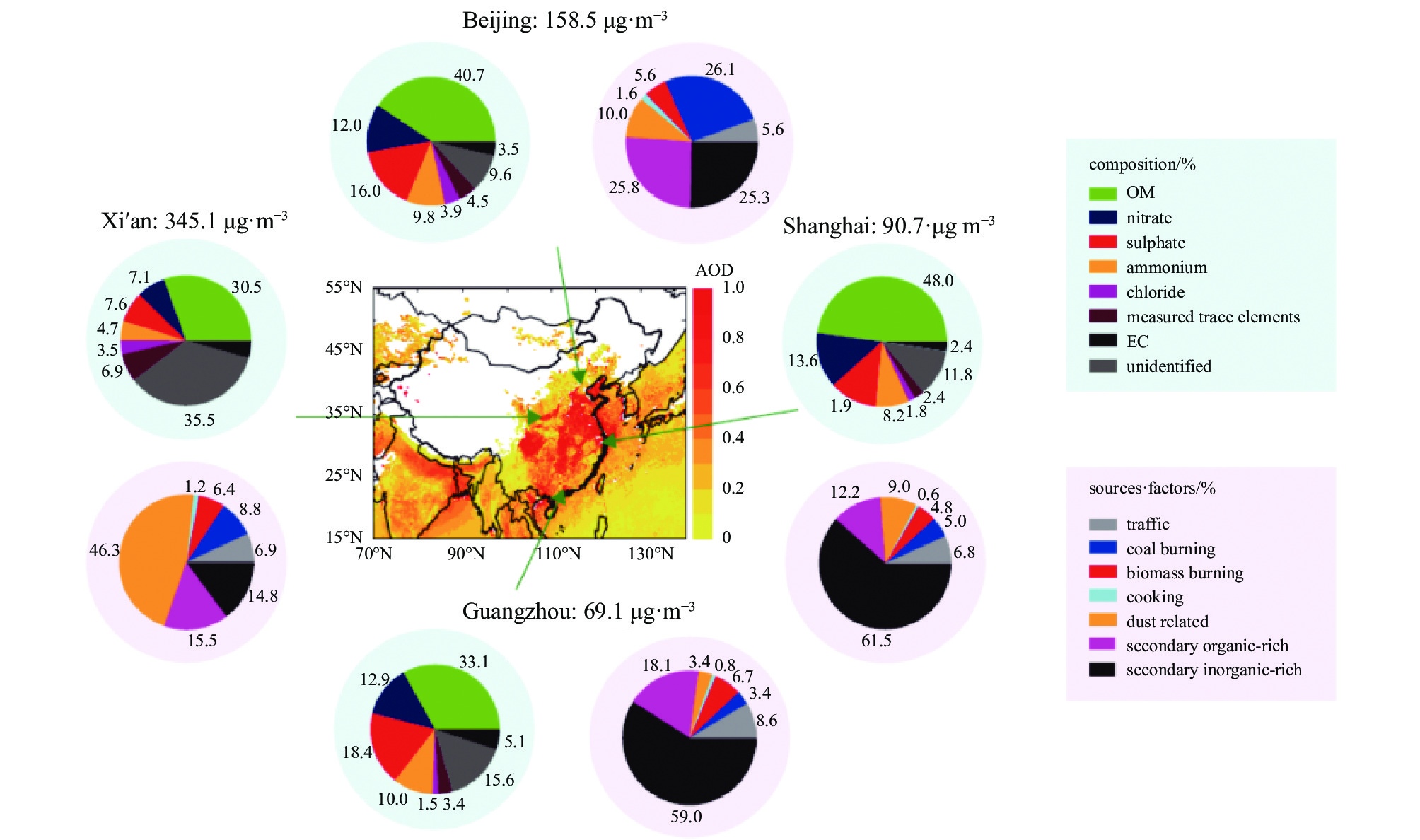
Huang Rujin, Zhang Yanlin, Bozzetti C, et al. High secondary aerosol contribution to particulate pollution during haze events in China[J]. Nature, 2014, 514(7521): 218-222. doi: 10.1038/nature13774
|
| [120] |
Yao Lei, Garmash O, Bianchi F, et al. Atmospheric new particle formation from sulfuric acid and amines in a Chinese megacity[J]. Science, 2018, 361(6399): 278-281. doi: 10.1126/science.aao4839
|
| [121] |
Johansson K O, Head-Gordon M P, Schrader P E, et al. Resonance-stabilized hydrocarbon-radical chain reactions may explain soot inception and growth[J]. Science, 2018, 361(6406): 997-1000. doi: 10.1126/science.aat3417
|
| [122] |
Taatjes C A, Welz O, Eskola A J, et al. Direct measurements of conformer-dependent reactivity of the criegee intermediate CH3CHOO[J]. Science, 2013, 340(6129): 177-180. doi: 10.1126/science.1234689
|
| [123] |
刘义鹤, 江洪. 5G通信新材料研究进展[J]. 新材料产业, 2019(8):51-53 doi: 10.19599/j.issn.1008-892x.2019.08.013
Liu Yihe, Jiang Hong. Research progress of new materials for 5G communication[J]. Advanced Materials Industry, 2019(8): 51-53 doi: 10.19599/j.issn.1008-892x.2019.08.013
|
| [124] |
Manaila-Maximean D. Effective permittivity of a multi-phase system: nanoparticle-doped polymer-dispersed liquid crystal films[J]. Molecules, 2021, 26: 1441. doi: 10.3390/molecules26051441
|
| [125] |
师文钊, 刘瑾姝, 邢建伟, 等. 聚乙烯醇基相变复合材料研究进展[J]. 中国材料进展, 2020, 39(3):234-242 doi: 10.7502/j.issn.1674-3962-201902015
Shi Wenzhao, Liu Jinshu, Xing Jianwei, et al. Research progress on polyvinyl alcohol-based phase change composites[J]. Materials China, 2020, 39(3): 234-242 doi: 10.7502/j.issn.1674-3962-201902015
|
| [126] |
Hong G, Gan Xuemin, Leonhardt C, et al. A brief history of OLEDs-emitter development and industry milestones[J]. Advanced Materials, 2021, 33: 2005630. doi: 10.1002/adma.202005630
|
| [127] |
Senses E, Ansar S M, Kitchens C L, et al. Small particle driven chain disentanglements in polymer nanocomposites[J]. Physical Review Letters, 2017, 118: 147801. doi: 10.1103/PhysRevLett.118.147801
|
| [128] |
Wang Maoyu, Árnadóttir L, Xu Z J, et al. In situ X-ray absorption spectroscopy studies of nanoscale electrocatalysts[J]. Nano-Micro Letters, 2019, 11: 47. doi: 10.1007/s40820-019-0277-x
|
| [129] |
刘涛, 纪军, 卫海桥, 等. 先进发动机燃烧基础研究的进展和关键科学问题[J]. 中国科学基金, 2014, 28(1):20-25 doi: 10.16262/j.cnki.1000-8217.2014.01.004
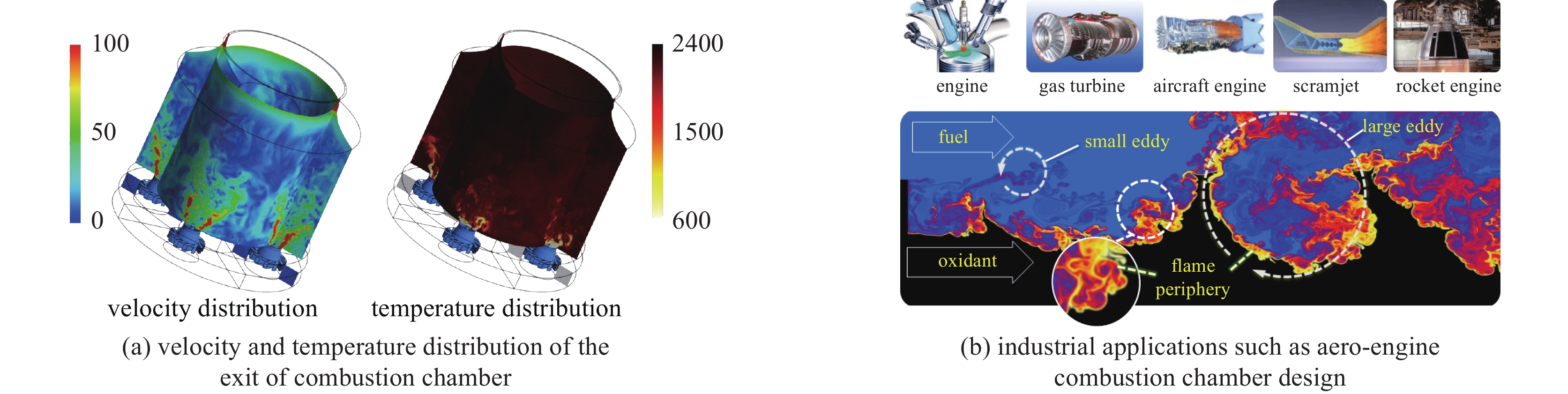
Liu Tao, Ji Jun, Wei Haiqiao, et al. Progress and key scientific issues on advanced engine combustion research—summary of the 92nd Shuangqing forum of NFSC[J]. Bulletin of National Natural Science Foundation of China, 2014, 28(1): 20-25 doi: 10.16262/j.cnki.1000-8217.2014.01.004
|
| [130] |
Dryer F L. Chemical kinetic and combustion characteristics of transportation fuels[J]. Proceedings of the Combustion Institute, 2015, 35(1): 117-144. doi: 10.1016/j.proci.2014.09.008
|
| [131] |
Qi Fei. Combustion chemistry probed by synchrotron VUV photoionization mass spectrometry[J]. Proceedings of the Combustion Institute, 2013, 34(1): 33-63. doi: 10.1016/j.proci.2012.09.002
|
| [132] |
Johansson K O, Dillstrom T, Monti M, et al. Formation and emission of large furans and oxygenated hydrocarbons from flames[J]. Proceedings of the National Academy of Sciences of the United States of America, 2016, 113(30): 8374-8379. doi: 10.1073/pnas.1604772113
|
| [133] |
Battin-Leclerc F, Herbinet O, Glaude P A, et al. Experimental confirmation of the low-temperature oxidation scheme of alkanes[J]. Angewandte Chemie International Edition, 2010, 49(18): 3169-3172. doi: 10.1002/anie.200906850
|
| [134] |
Yang Bin, Oßwald P, Li Yuyang, et al. Identification of combustion intermediates in isomeric fuel-rich premixed butanol–oxygen flames at low pressure[J]. Combustion and Flame, 2007, 148(4): 198-209. doi: 10.1016/j.combustflame.2006.12.001
|
| [135] |
Taatjes C A, Hansen N, McIlroy A, et al. Enols are common intermediates in hydrocarbon oxidation[J]. Science, 2005, 308(5730): 1887-1889. doi: 10.1126/science.1112532
|
| [136] |
Qi F, Li Y Y, et al. The second prize in National Natural Science Awards. 2018.
|
Figures(17)


 DownLoad: