

Single energy X-ray source for calibration of X-ray detectors
-
摘要: 为提高X射线探测器的标定精度,在荧光X射线源的基础上,提出在荧光X射线出射通道设置滤光片的方法提高X射线纯度。通过蒙特卡罗建立仿真模型,分析了辐射体发生K层光电效应的概率与原子序数的关系,并得到荧光强度和纯度随滤光片厚度的变化曲线。在大气环境下,采用硅漂移半导体探测器测试了荧光X射线源的能谱分布和光子流量,分析X射线管管电压对光子流量和荧光纯度的影响。在辐射体材料为铜,滤光片(镍)厚度为0 μm、10 μm和30 μm时,测得的荧光X射线纯度分别为75.61%、85.38%和84.25%,光子流量分别为3425 phs/s、2023 phs/s和1192 phs/s,确认了滤光片厚度对荧光X射线纯度和强度的影响,为解决荧光X射线光源单色性不足难以对X射线探测器进行高精度标定的问题提供了方向。Abstract: To improve the calibration accuracy of X-ray detectors, this paper presents a method of placing filters in fluorescent X-ray emission channels to improve the purity of X-rays. Monte Carlo simulation model was established to analyze the relationship between the probability of photoelectric effect in K layer and the atomic number, and the curve of fluorescence intensity and purity with filter thickness was obtained. In atmospheric environment, the energy spectrum distribution and photon flux of fluorescent X-ray source were measured by silicon drift semiconductor detector, and the effect of X-ray tube voltage on photon flux and fluorescence purity was analyzed. When the radiator material is copper and the thickness of the filter (nickel) is 0 μm, 10 μm and 30 μm, the purity of fluorescence X-ray measured is 75.61%, 85.38% and 84.25%, and the photon flux is 3425 phs/s, 2023 phs/s and 1192 phs/s, respectively. The influence of filter thickness on the purity and intensity of fluorescent X-ray is confirmed, which provides a direction for solving the problem that it is difficult to calibrate X-ray detectors with high accuracy due to the lack of monochromatism of fluorescent X-ray light source.
-
Key words:
- fluorescent X-ray source /
- filter /
- calibration of detector
-
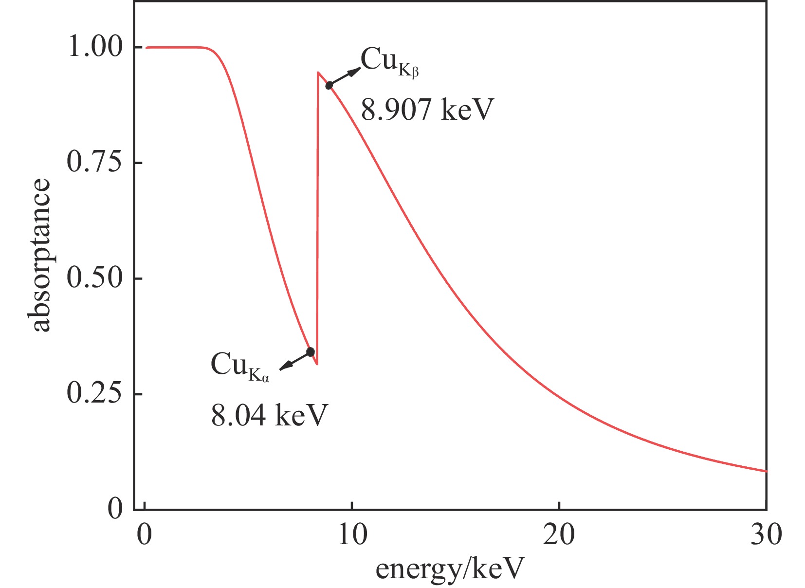
图 3 厚度为10 μm的Ni片的吸收率变化曲线
Figure 3. Change curve of absorptivity of Ni sheet with thickness of 10 μm
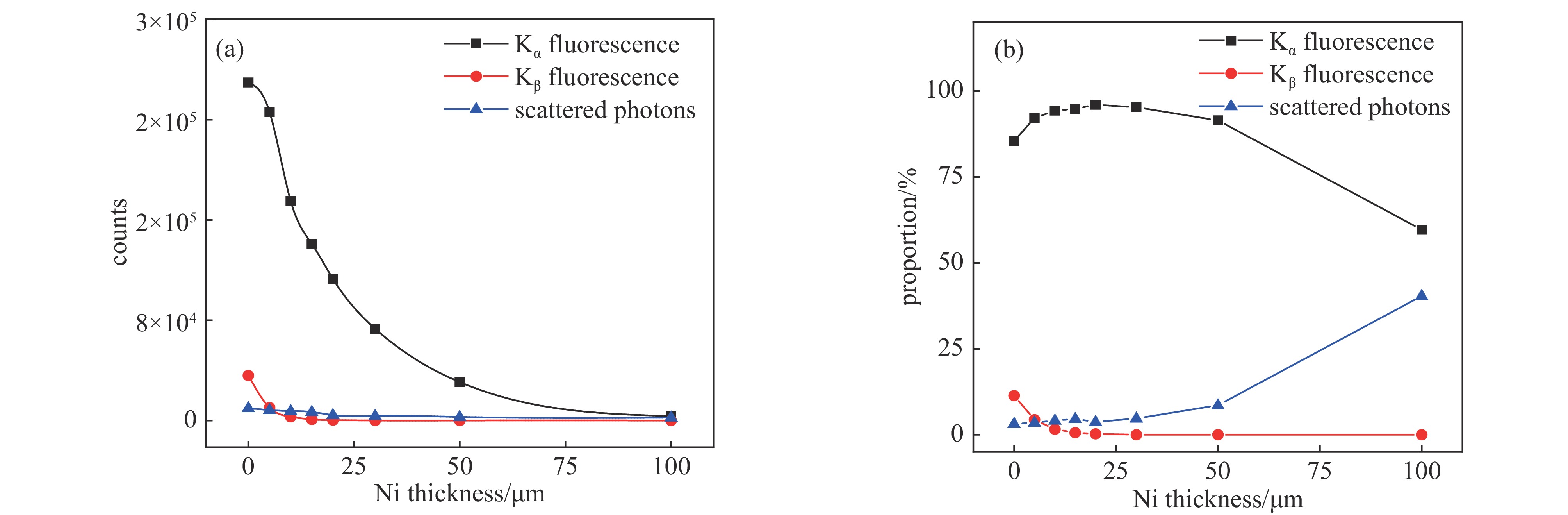
图 4 辐射体Cu的Kα、Kβ和散射光子的强度和占比随Ni片厚度变化曲线
Figure 4. Variation curve of intensity and proportion of the radiator Cu with the thickness of the Ni sheet
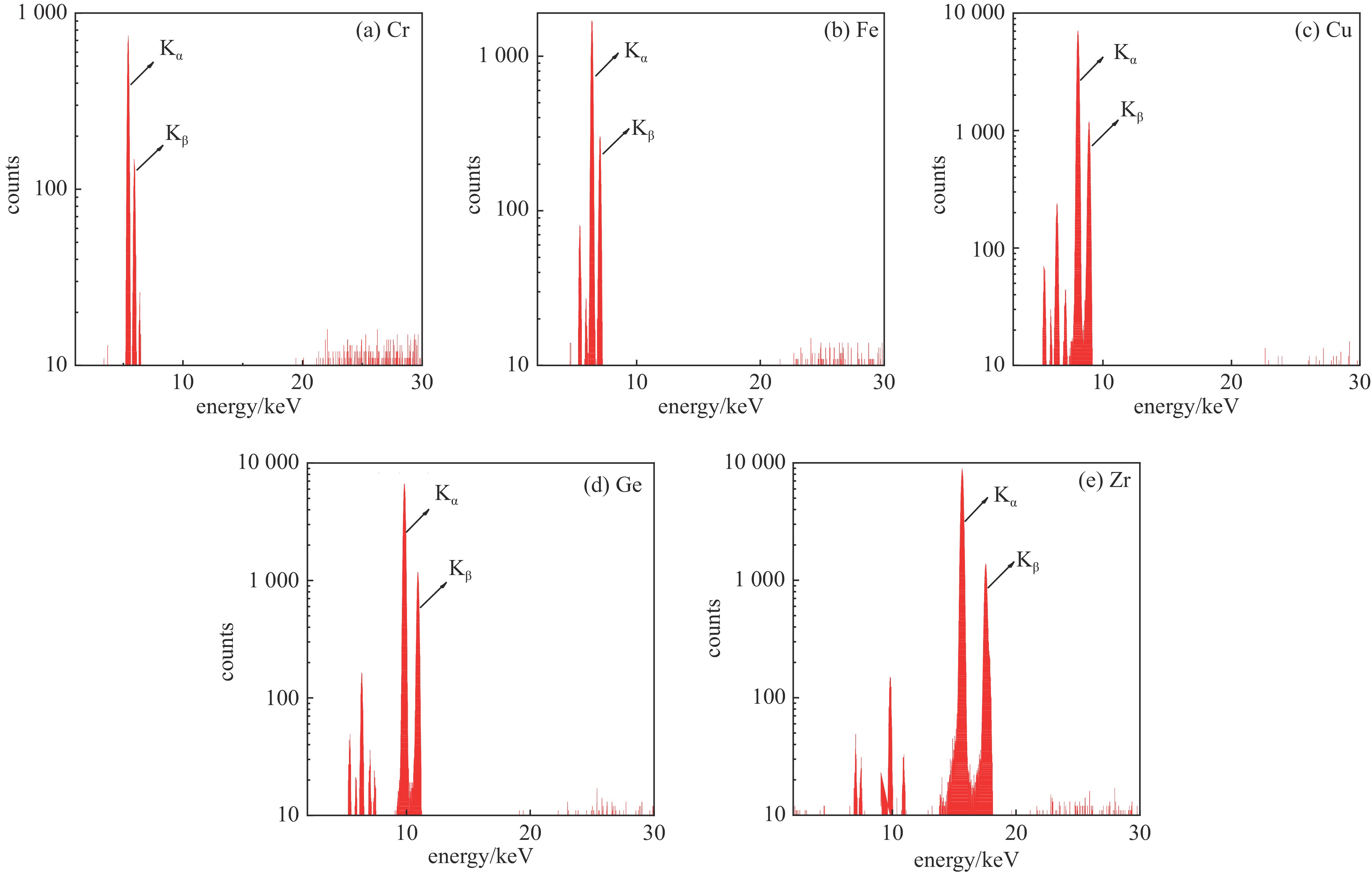
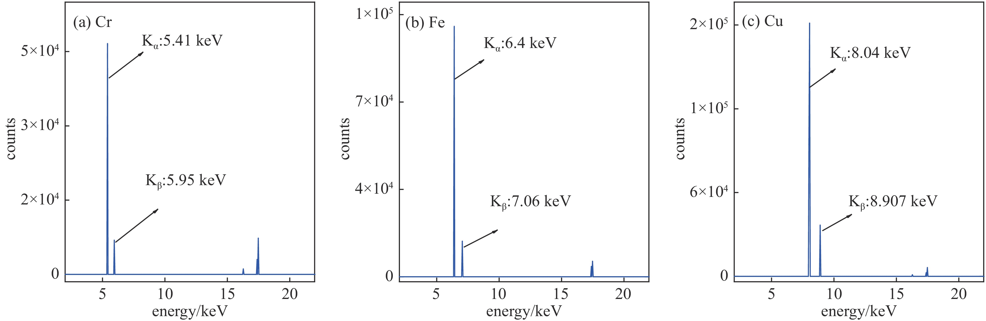
图 6 不同辐射体材料的荧光X射线能谱
Figure 6. Fluorescence X-ray energy spectrum of different radiation materials
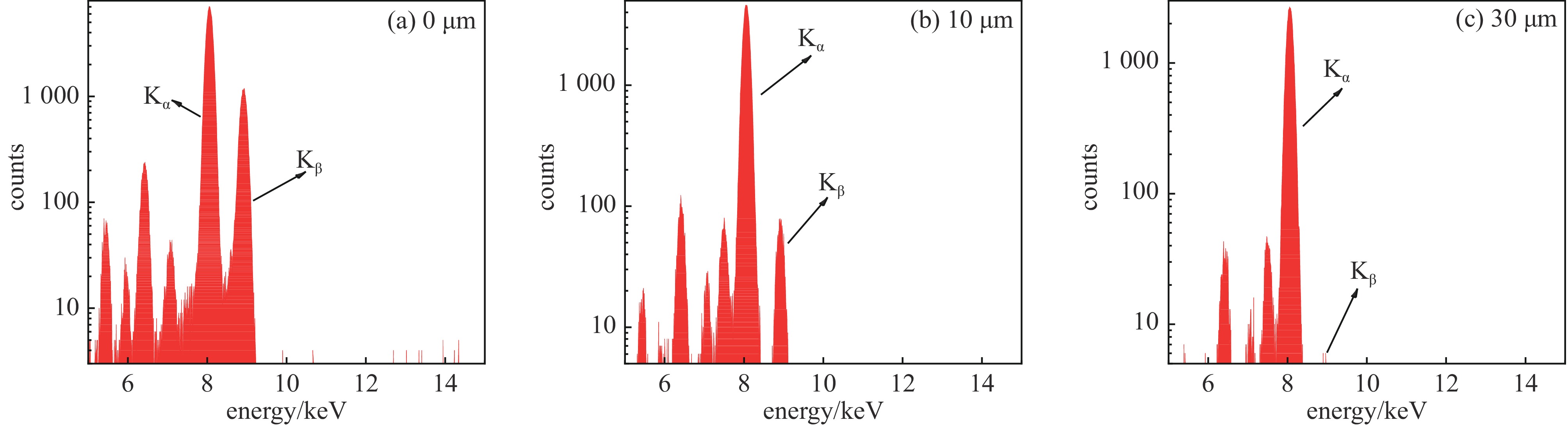
图 7 不同Ni片厚度辐射体Cu的荧光X射线能谱
Figure 7. Fluorescence X-ray energy spectrum of Cu in radiators with different Ni thickness
表 1 不同辐射体的荧光X射线源能谱仿真数据
Table 1. Spectral simulation data of fluorescence X-ray sources with different radiators
material Kα photons Kα proportion/% K photons K proportion/% scattered photons scattered photon proportion/% Cr 46622 79.791 53529 82.426 64941 17.574 Fe 100289 80.270 114567 91.698 10373 8.302 Cu 252727 85.466 286400 96.853 9304 3.147  下载: 导出CSV
表 2 测得的不同辐射体材料的能谱数据
Table 2. Measured spectral data of different radiation materials
material $ {\rm{K}}_ {\text{α}} $ photons $ {\rm{K}}_ {\text{α}}$ proportion/% K photons K proportion/% scattered photons scattered photon proportion/% Cr 15410 45.72 18273 54.215 15432 45.785 Fe 39177 64.017 46347 75.733 14850 24.267 Cu 155442 75.642 183030 89.067 22466 10.933 Ge 177050 77.485 208868 91.410 19627 8.59 Zr 298287 79.044 351258 93.081 25733 6.819
下载: 导出CSV
表 3 不同
$ {\mathit{V}}_{\rm{a}} $ 下测得的辐射体Cu的光子流量数据Table 3. Photon flux data of radiator Cu measured under different
$ {\mathit{V}}_{\rm{a}} $ Ia/μA Va/kV Kα counting rate/(phs·s−1) Kα proportion/% scattered photon proportion/% 200 20 45 79.147 7.856 200 25 231 78.247 8.360 200 30 600 77.253 9.182 200 35 1007 76.568 9.734 200 40 1509 76.013 10.437 200 45 2047 75.815 10.491 200 50 2589 75.608 10.932
下载: 导出CSV
-
[1] Li Tipei, Xiong Shaolin, Zhang Shuangnan, et al. Insight-HXMT observations of the first binary neutron star merger GW170817[J]. Science China Physics, Mechanics & Astronomy, 2018, 61: 031011. [2] Tuo Youli, Ge Mingyu, Song Liming, et al. Insight-HXMT observations of the Crab pulsar[J]. Research in Astronomy and Astrophysics, 2019, 19: 087. doi: 10.1088/1674-4527/19/6/87 [3] Zhang Dali, Li Xinqiao, Xiong Shaolin, et al. Energy response of GECAM gamma-ray detector based on LaBr3: Ce and SiPM array[J]. Nuclear Instruments and Methods in Physics Research Section A: Accelerators, Spectrometers, Detectors and Associated Equipment, 2019, 921: 8-13. [4] Wang Shen, Guo Jianhua, Zhang Yan, et al. High-resolution pixelated CdZnTe detector prototype system for solar hard X-ray imager[J]. Nuclear Science and Techniques, 2019, 30: 42. doi: 10.1007/s41365-019-0571-9 [5] Dong Yongwei, Wu Bobing, Li Yanguo, et al. SVOM gamma ray monitor[J]. Science China Physics, Mechanics and Astronomy, 2010, 53(1): 40-42. [6] Götz D, Paul J, Basa S, et al. SVOM: a new mission for gamma-ray burst studies[J]. AIP Conference Proceedings, 2009, 1133: 25-30. [7] Yuan Weimin, Zhang Chen, Chen Yong, et al. Einstein probe: exploring the ever-changing X-ray universe[J]. SCIENTIA SINICA Physica, Mechanica & Astronomica, 2018, 48: 039502. [8] Zhang Shuangnan, Santangelo A, Feroci M, et al. The enhanced X-ray timing and polarimetry mission—eXTP[J]. Science China Physics, Mechanics & Astronomy, 2019, 62: 29502. [9] Guo Siming, Jiang Zheng, Wu Jinjie, et al. Research on a tunable monochromatic X-rays source in (5~40) keV[J]. Applied Radiation and Isotopes, 2022, 181: 110096. doi: 10.1016/j.apradiso.2022.110096 [10] Kobayashi K, Yabashi M, Takata Y, et al. High resolution-high energy X-ray photoelectron spectroscopy using third-generation synchrotron radiation source, and its application to Si-high k insulator systems[J]. Applied Physics Letters, 2003, 83(5): 1005-1007. doi: 10.1063/1.1595714 [11] Zhou Xu, Li Xinqiao, Xie Yaning, et al. Introduction to a calibration facility for hard X-ray detectors[J]. Experimental Astronomy, 2014, 38(3): 433-441. doi: 10.1007/s10686-014-9393-2 [12] Gambaccini M, Tuffanelli A, Taibi A, et al. Bragg-diffraction-based quasi-monochromatic source for mammography using mosaic crystals[C]//Proceedings of SPIE 3770, Medical Applications of Penetrating Radiation. 1999: 174-184. [13] Csete I. Production of fluorescent X-rays from 8 to 100 keV[J]. International Journal of Radiation Applications and Instrumentation. Part A. Applied Radiation and Isotopes, 1992, 43(6): 767-776. doi: 10.1016/0883-2889(92)90240-F [14] 代锦飞, 赵宝升, 盛立志, 等. 标定脉冲星导航探测器的荧光X射线光源[J]. 物理学报, 2015, 64:149071 doi: 10.7498/aps.64.149701Dai Jinfei, Zhao Baosheng, Sheng Lizhi, et al. Ffluorescence X-ray source used for calibrating the detector of X-ray navigation[J]. Acta Physica Sinica, 2015, 64: 149071 doi: 10.7498/aps.64.149701 [15] 祝宇轩, 王于仨, 陈勇, 等. 用于软X射线探测器标定的X射线二次多靶源[J]. 核技术, 2021, 44:050402 doi: 10.11889/j.0253-3219.2021.hjs.44.050402Zhu Yuxuan, Wang Yusa, Chen Yong, et al. X-ray secondary multiple target sources for calibration of soft X-ray detectors[J]. Nuclear Techniques, 2021, 44: 050402 doi: 10.11889/j.0253-3219.2021.hjs.44.050402 [16] Bambynek W, Crasemann B, Fink R W, et al. X-ray fluorescence yields, Auger, and Coster-Kronig transition probabilities[J]. Reviews of Modern Physics, 1972, 44(4): 716-813. doi: 10.1103/RevModPhys.44.716 [17] Ménesguen Y, Lépy M C. Mass attenuation coefficients in the range 3.8≤E≤11 keV, K fluorescence yield and Kβ/Kα relative X-ray emission rate for Ti, V, Fe, Co, Ni, Cu and Zn measured with a tunable monochromatic X-ray source[J]. Nuclear Instruments and Methods in Physics Research Section B: Beam Interactions with Materials and Atoms, 2010, 268(16): 2477-2486. doi: 10.1016/j.nimb.2010.05.044 [18] 梁敬魁. 粉末衍射法测定晶体结构-上册: X射线衍射结构晶体学基础[M]. 2版. 北京: 科学出版社, 2011Liang Jingkui. Determination of crystal structure by powder method (Volume 1)[M]. 2nd ed. Beijing: Science Press, 2011 [19] 盛立志, 赵宝升, 吴建军, 等. X射线脉冲星导航系统模拟光源的研究[J]. 物理学报, 2013, 62:129702 doi: 10.7498/aps.62.129702Sheng Lizhi, Zhao Baosheng, Wu Jianjun, et al. Research of X-ray pulsar navigation simulation source[J]. Acta Physica Sinica, 2013, 62: 129702 doi: 10.7498/aps.62.129702 [20] Storm E. Bremsstrahlung-induced K-fluorescent radiation[J]. Journal of Applied Physics, 1976, 47(7): 3060-3070. doi: 10.1063/1.323053 -


 点击查看大图
计量
- 文章访问数: 947
- HTML全文浏览量: 284
- PDF下载量: 85
- 被引次数: 0
