

Surface-enhanced Raman effect of new coronavirus S protein in gold nanoparticles
-
摘要: 表面增强拉曼光谱技术因其高灵敏度、操作简单、快速检测等优点,被广泛用于病毒检测方面。国内外的病毒拉曼检测研究主要集中在检测病毒核酸以及组成核酸的各种碱基的表面增强拉曼光谱(SERS),但少见对病毒蛋白的SERS检测。以新型冠状病毒(SARS-CoV-2)的S蛋白为检测对象,采用无标记SERS检测方法,对比SARS-CoV-2固态、饱和液态S蛋白的普通拉曼光谱和选用40 nm金纳米粒子为基底的SARS-CoV-2低浓度S蛋白SERS光谱。结果表明,以40 nm金纳米粒子为基底,采用SERS技术检测SARS-CoV-2的S蛋白是完全可行的。SARS-CoV-2的S蛋白分子中的羧基与金纳米粒子发生了分子增强,氨基与金纳米粒子发生了电磁增强,从而使得SARS-CoV-2的S蛋白拉曼效应得到了增强,并使得峰位发生一定移动。实验获得了较好的SARS-CoV-2低浓度S蛋白SERS光谱,为建立敏感、特异、快速的SARS-CoV-2检测新技术提供了一种方法。
-
关键词:
- 表面增强拉曼光谱技术 /
- 新型冠状病毒 /
- 金纳米粒子 /
- 蛋白质 /
- 相互作用
Abstract: Surface-enhanced Raman spectroscopy (SERS) technology has been widely used in viral molecular detection due to its high sensitivity, simple operation and rapid detection. The research of virus detection by Raman technology at home and abroad mainly focuses on the detection of the SERS spectrum of viral nucleic acids and various bases that make up the nucleic acids, and detection of viral proteins is rare. In this paper, the S protein of the new coronavirus (SARS-CoV-2) is used as the detection object, and with the label-free SERS detection method, the ordinary Raman spectra of solid and saturated liquid S protein of the SARS-CoV-2 and the SERS spectra of the low-concentration S protein of SARS-CoV-2 on the substrate of gold nanoparticles with a size of 40 nm are compared. The results show that it is completely feasible to use SERS technology to detect the S protein of SARS-CoV-2 on the substrate of 40 nm gold nanoparticles. The carboxyl groups in the S protein molecule of SARS-CoV-2 and gold nanoparticles are molecularly enhanced, and the amino groups and gold nanoparticles are electromagnetically enhanced, so that the Raman effect of the S protein of the SARS-CoV-2 is enhanced and the peak position is moved to a certain extent. The experiments obtained relatively good SERS spectra of the low-concentration S protein of SARS-CoV-2, which provides a method for the establishment of a sensitive, specific and rapid detection technology for the S protein of the SARS-CoV-2. -
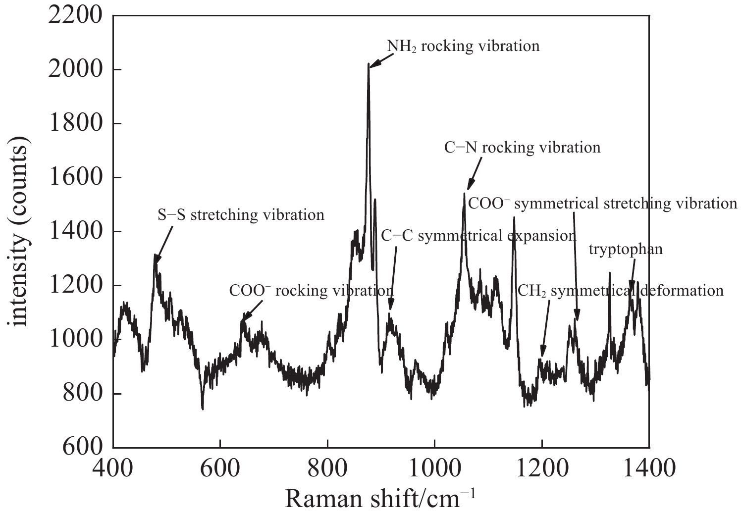
图 1 SARS-CoV-2固态S蛋白的拉曼光谱
Figure 1. Raman spectrum of the S protein of SARS-CoV-2 in solid state
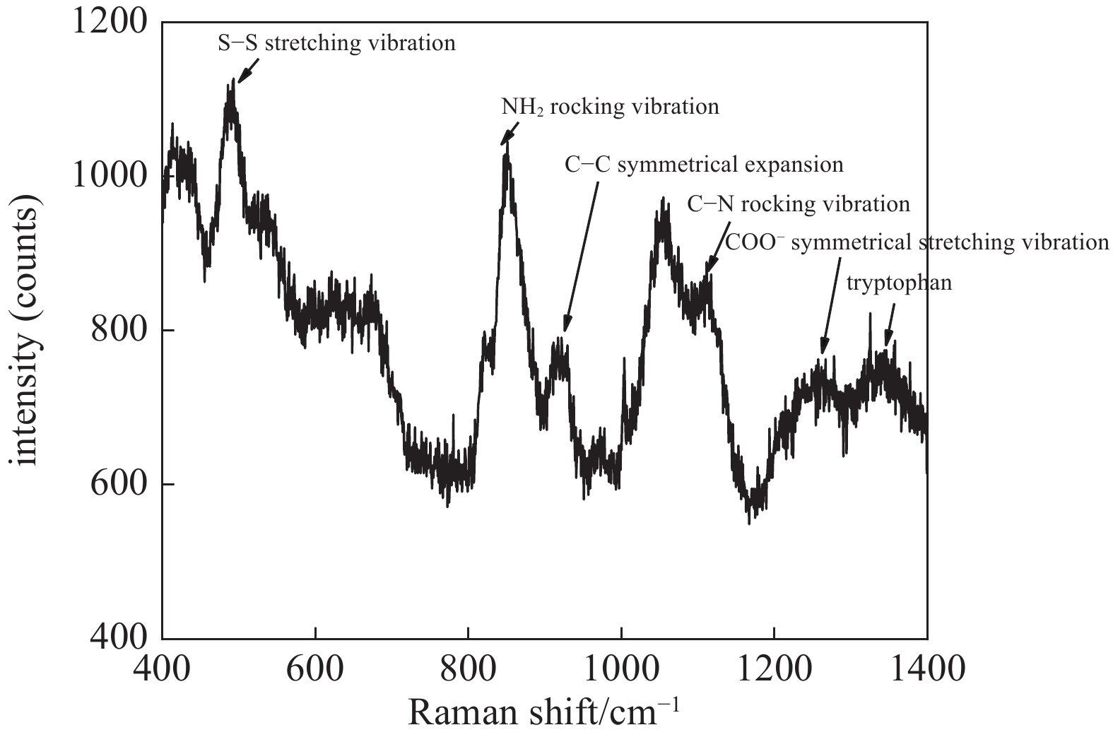
图 2 SARS-CoV-2饱和液态S蛋白的拉曼光谱
Figure 2. Raman spectrum of the S protein of SARS-CoV-2 in saturated liquid state
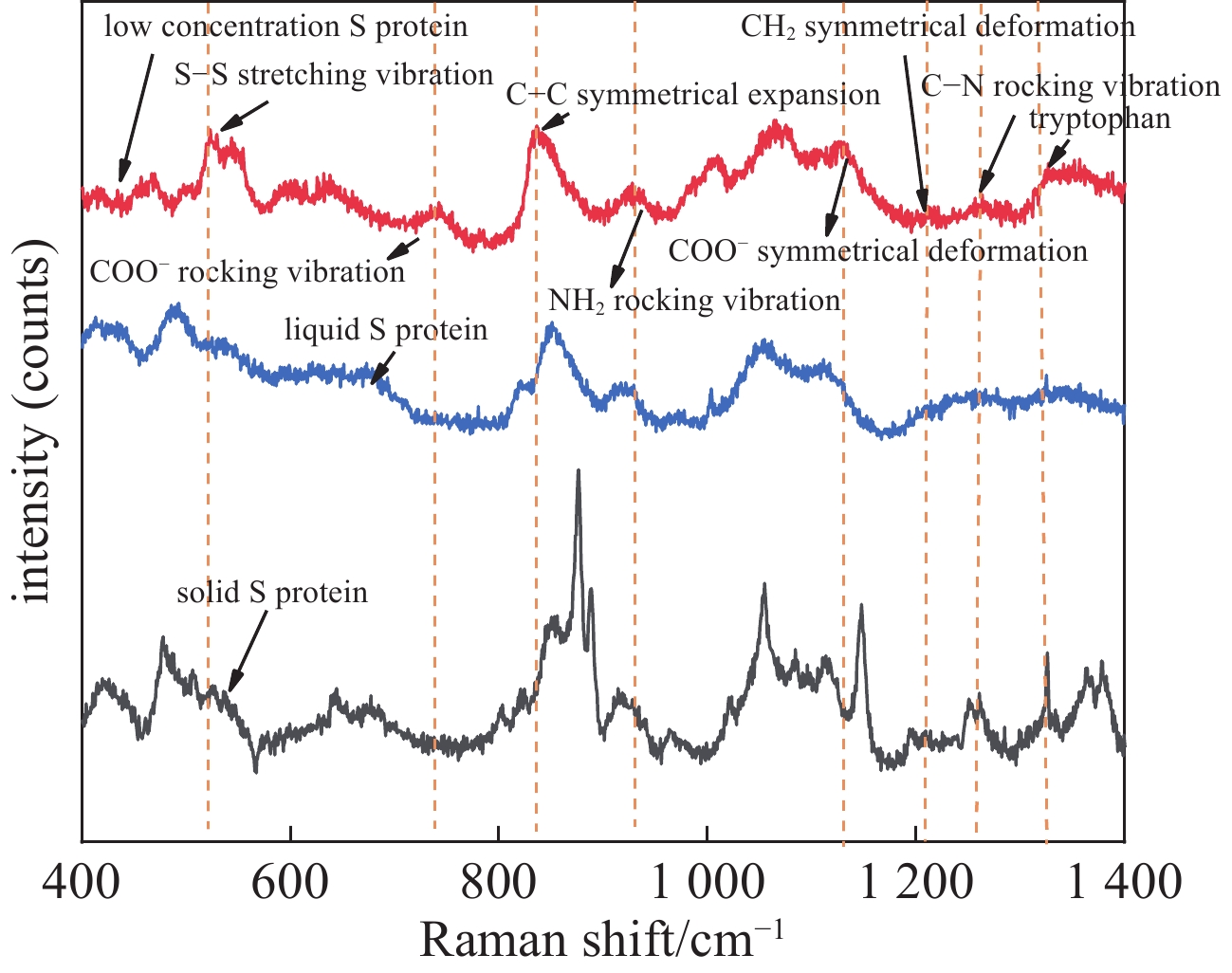
图 3 SARS-CoV-2 S蛋白的常规拉曼光谱和SERS光谱
Figure 3. Conventional Raman spectrum and SERS spectra of the S protein of SARS-CoV-2
表 1 SARS-CoV-2的S蛋白的主要峰位(cm−1)与归属[14−16]
Table 1. Raman peaks (cm−1) and their assignment of the S protein of SARS-CoV-2[14−16]
peak of pure solid
SARS-CoV-2/cm−1peak of SARS-CoV-2 in
saturated liquid state/cm−1SERS of
SARS-CoV-2/cm−1assignment of
the bands537.7 524.7 523.3 S−S stretching vibration 679.1 / 742.6 COO− rocking vibration 851.6 850.7 836.2 NH2 rocking vibration 915.3 917.1 927.6 C−C symmetrical expansion 1111.1 1111.4 1126.3 C−N rocking vibration 1250.9 / 1211.5 CH2 symmetrical deformation 1326.4 1257.2 1263.5 COO− symmetrical stretching vibration 1362.7 1343.6 1357.3 tryptophan  下载: 导出CSV
-
[1] Ceraolo C, Giorgi F M. Genomic variance of the 2019 nCoV coronavirus[J]. Journal of Medical Virology, 2020, 92(5): 522-528. doi: 10.1002/jmv.25700 [2] 朱宁. 加强国际合作 携手抗击疫情[J]. 浙江经济期刊, 2020(4):76. (Zhu Ning. Strengthen international cooperation to fight the epidemic[J]. Zhejiang Economy, 2020(4): 76 [3] Coronavirus disease 2019 (COVID-19) situation report-65[EB/OL].https://www.who.int/docs/default-source/coronaviruse/situation-reports/20200325-sitrep-65-covid-19.pdf?sfvrsn=ce13061b_2. [4] He Yi, Yang Xia, Yuan Ruo, et al. Switchable target-responsive 3D DNA hydrogels as a signal amplification strategy combining with SERS technique for ultrasensitive detection of miRNA 155[J]. Analytical Chemistry, 2017, 89(16): 8538-8544. doi: 10.1021/acs.analchem.7b02321 [5] 王越珉, 雷喜梅, 邬丽, 等. 新型冠状病毒及其检测方法研究进展[J]. 中国计量大学学报, 2020, 31(1):1-7. (Wang Yuemin, Lei Ximei, Wu Li, et al. A review of severe acute respiratory syndrome coronavirus 2 and its detecting methods[J]. Journal of China University of Metrology, 2020, 31(1): 1-7 doi: 10.3969/j.issn.2096-2835.2020.01.001 [6] 李晓楠. 一种新型冠状病毒S蛋白和N蛋白联合检测胶体金试纸条及其制备方法和用途: 202010851052.5[P]. 2021-02-26. [7] Lipkowski J, Stolberg L, Yang Dongfang, et al. Molecular adsorption at metal electrodes[J]. Electrochimica Acta, 1994, 39(8/9): 1045-1056. [8] 柯惟中, 吴缄中. 氨基酸在银胶溶液中的表面增强拉曼效应[J]. 光谱学与光谱分析, 2004, 24(5):551-553. (Ke Weizhong, Wu Jianzhong. Surface-Enhanced Raman Scattering (SERS) of Amino acids on silver colloid[J]. Spectroscopy and Spectral Analysis, 2004, 24(5): 551-553 doi: 10.3321/j.issn:1000-0593.2004.05.010 [9] Panikkanvalappil SP, Mackey MA, El-Sayed MA. Probing the unique dehydration-induced structural modifications in cancer cell DNA using surface enhanced Raman spectroscopy[J]. Journal of the American Chemical Society, 2013, 135(12): 4815-4821. doi: 10.1021/ja400187b [10] Li Xiaoxiao, Ye Sujuan, Luo Xiliang. Sensitive SERS detection of miRNA via enzyme-free DNA machine signal amplification[J]. Chemical Communications, 2016, 52(67): 10269-10272. doi: 10.1039/C6CC04391G [11] 周民杰. 一种基于增强拉曼光谱和神经网络的新型冠状病毒检测方法及系统: 202110006417.9[P]. 2021-05-14.Zhou Minjie. New method and system for SARS-CoV-2 detection based on enhanced Raman spectrum and neural network: 202110006417.9[P]. 2021-05-14. [12] 黄景林, 周民杰, 乐玮, 等. 表面增强拉曼光谱技术检测新型冠状病毒刺突蛋白[J]. 强激光与粒子束, 2020, 32:069001. (Huang Jinglin, Zhou Mingjie, Le Wei, et al. Detection of spike protein of SARS-CoV-2 by surface enhanced Raman spectroscopy[J]. High Power Laser and Particle Beams, 2020, 32: 069001 doi: 10.11884/HPLPB202032.200145 [13] 王晓辉, 徐涛涛, 黄轶群, 等. 表面增强拉曼光谱在食源性致病微生物检测中的应用研究[J]. 光谱学与光谱分析, 2019, 39(1):123-129. (Wang Xiaohui, Xu Taotao, Huang Yiqun, et al. Application of surface-enhanced Raman spectroscopy for foodborne pathogens detection[J]. Spectroscopy and Spectral Analysis, 2019, 39(1): 123-129 [14] Doering W E, Nie Shuming. Single-molecule and single-nanoparticle SERS: examining the roles of surface active sites and chemical enhancement[J]. The Journal of Physical Chemistry B, 2002, 106(2): 311-317. doi: 10.1021/jp011730b [15] Stewart S, Fredericks P M. Surface-enhanced Raman spectroscopy of peptides and proteins adsorbed on an electrochemically prepared silver surface[J]. Spectrochimica Acta Part A:Molecular and Biomolecular Spectroscopy, 1999, 55(7/8): 1615-1640. [16] 胡国进, 余文玉. 生物分子的表面增强拉曼散射[J]. 江西教育学院学报(自然科学), 2002, 23(3):18-22. (Hu Guojin, Yu Wenyu. On the surface enhancement L-M scatter of biological molecule[J]. Journal of Jiangxi Institute of Education (Natural Sciences), 2002, 23(3): 18-22 [17] 张丹. 氨基酸的表面增强拉曼光谱研究[D]. 杭州: 浙江工业大学, 2006: 52-54Zhang Dan. The study of amino acid by surface-enhanced Raman scattering[D]. Hangzhou: Zhejiang University of Technology, 2006: 52-54 [18] 潘家来. 激光拉曼光谱在有机化学上的应用[M]. 北京: 化学工业出版社, 1986Pan Jialai. Application of laser Raman spectroscopy in organic chemistry[M]. Beijing: Chemical Industry Press, 1986 [19] 朱自莹, 顾仁熬, 陆天虹. 拉曼光谱在化学中的应用[M]. 沈阳: 东北大学出版社, 1998Zhu Ziying, Gu Ren’ao, Lu Tianhong. The application of Raman spectroscopy in chemistry[M]. Shenyang: Northeastern University Press, 1998 -

 点击查看大图
计量
- 文章访问数: 1689
- HTML全文浏览量: 667
- PDF下载量: 60
- 被引次数: 0
