

Micro-focus computed tomography for turbine blade based on all-optical bremsstrahlung source
-
摘要:
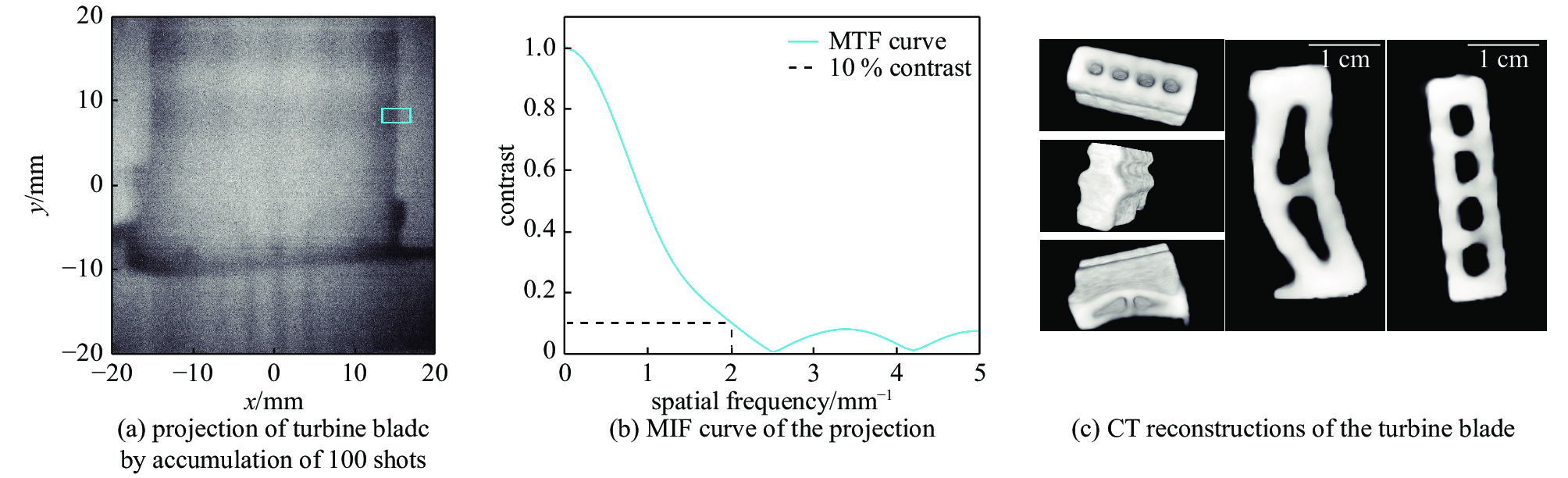
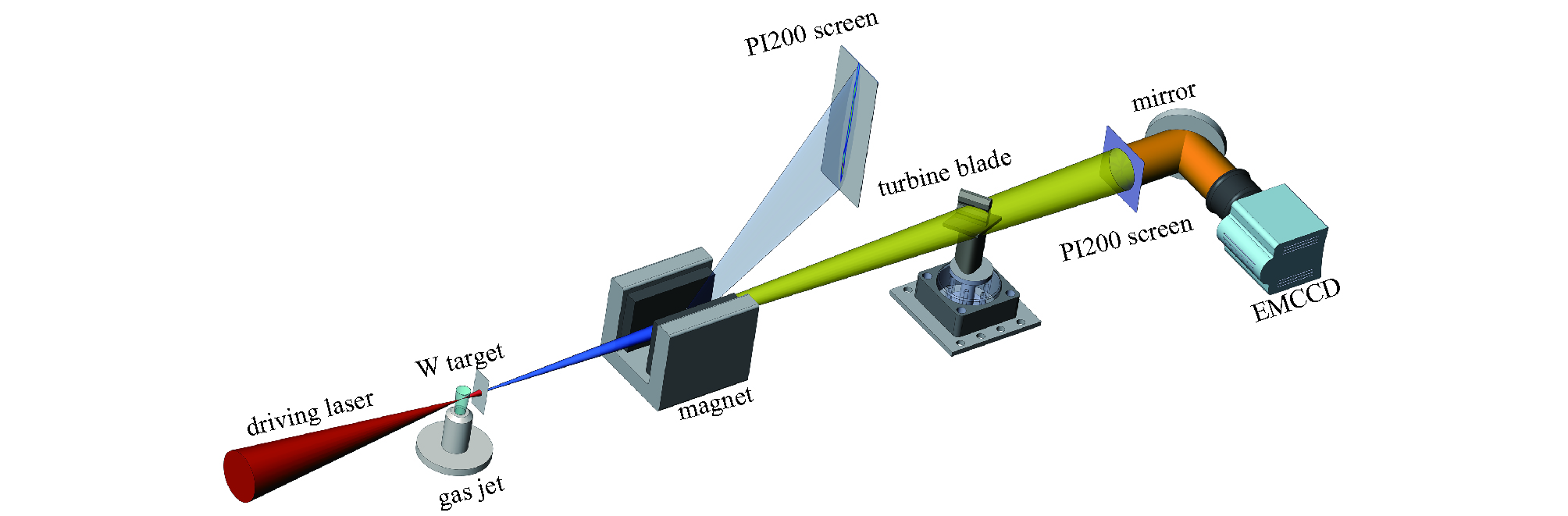
发展微焦点高能X射线源技术是实现高精度高能工业CT突破的关键,基于激光尾波加速驱动高能轫致辐射源开展了微焦点高能X射线源产生以及对涡轮叶片高能CT成像研究。利用一台20 TW钛蓝宝石超快超强激光器,通过电离注入的方式获得了(140±44)pC的高能电子束,并使用1.5 mm厚钨靶产生了累积源尺寸为25 μm的高能轫致辐射X射线。利用该微焦点高能X射线源,采用基于压缩感知的CT重建算法,在获取较少角度投影(31个角度)的情况下,获得了对涡轮叶片叶榫结构的CT重建。
Abstract:Computed tomography is a major technique for nondestructive detection of internal defects of dense materials and large-size devices. It is widely used in material science, railway, aerospace, national defense, military industry and other industries. At present, conventional high-energy industrial computed tomography system uses the X-ray source based on traditional thermionic RF electron gun, which can only provide millimeter source size, thus limiting its imaging spatial resolution. A high-energy micro-focus X-ray source is the key means to realize high-resolution high-energy industry computed tomography. As an emerging accelerator technology, the laser wakefield accelerator is a promising candidate for the micro-focus high-energy industrial computed tomography. This article reports experimental results of a micro-focus X-ray source based on laser wakefield acceleration and a computed tomography for a turbine blade. Using a 20 TW Ti: sapphire laser system, an electron beam with a charge of (140±44) pC is generated through ionization-induced injection, and then an all-optical bremsstrahlung X-ray source with an accumulated source size of 25 μm is obtained by using a 1.5 mm tungsten target. Using this source, a preliminary compressed-sensing-based computed tomography for a turbine blade is performed.
-
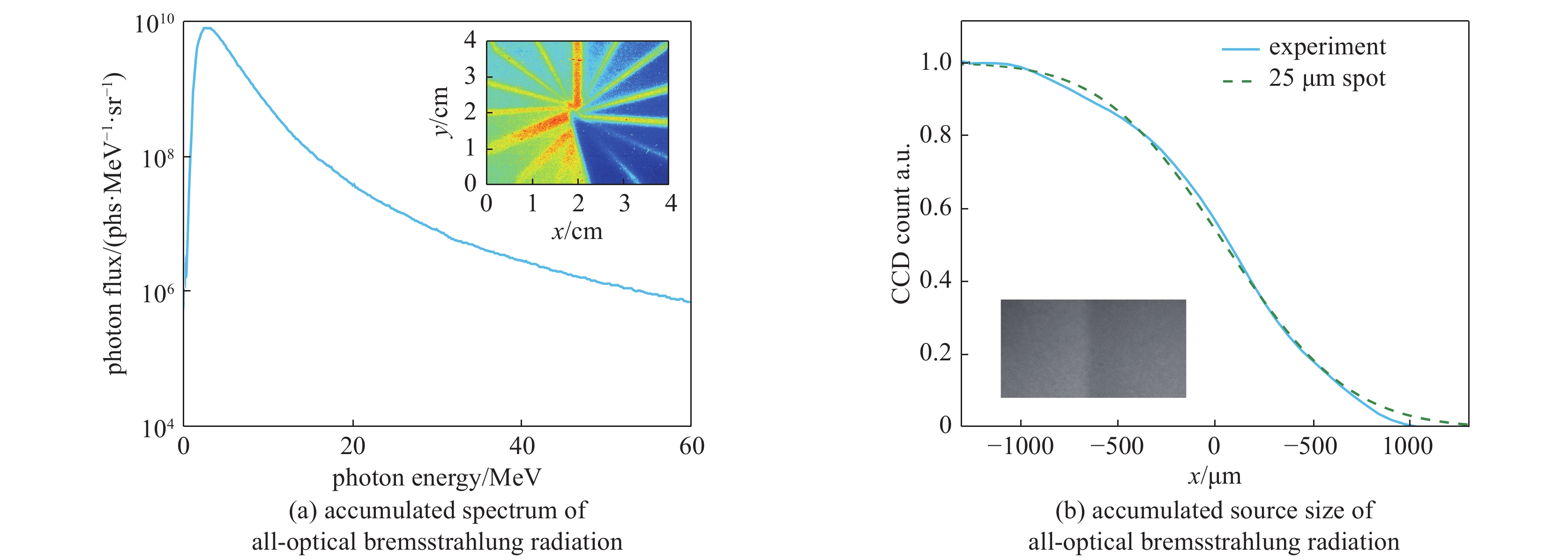
图 3 连续100发全光轫致辐射累积能谱以及累积源尺寸测量结果
Figure 3. Measured accumulated spectrum and accumulated source size of all-optical bremsstrahlung radiation for 100 consecutive shots
-
[1] Yamaguchi K, Ohtake Y, SuzukiH, et al. Dimensional measurement of nuclear fuel pellets using high energy X-ray CT[C]//Proc of Non-Destructive Testing. 2016, 7: 1-7. [2] Tajima T, Dawson J M. Laser electron accelerator[J]. Physical Review Letters, 1979, 43(4): 267-270. doi: 10.1103/PhysRevLett.43.267 [3] Lu W, Huang C, Zhou M, et al. Nonlinear theory for relativistic plasma wakefields in the blowout regime[J]. Physical Review Letters, 2006, 96: 165002. doi: 10.1103/PhysRevLett.96.165002 [4] Leemans W P, Gonsalves A J, Mao H S, et al. Multi-GeV electron beams from capillary-discharge-guided subpetawatt laser pulses in the self-trapping regime[J]. Physical Review Letters, 2014, 113: 245002. doi: 10.1103/PhysRevLett.113.245002 [5] 陈民, 刘峰, 李博原, 等. 激光等离子体尾波加速器的发展和展望[J]. 强激光与粒子束, 2020, 32:092001. (Chen Min, Liu Feng, Li Boyuan, et al. Development and prospect of laser plasma wakefield accelerator[J]. High Power Laser and Particle Beams, 2020, 32: 092001 doi: 10.11884/HPLPB202032.200174 [6] 盛政明, 陈民, 翁苏明, 等. 超短超强激光驱动新型粒子加速器: 机遇和挑战[J]. 物理, 2018, 47(12):753-762. (Sheng Zhengming, Chen Min, Weng Suming, et al. Novel particle accelerators driven by ultrashort and ultraintense lasers: opportunities and challenges[J]. Phyisics, 2018, 47(12): 753-762 doi: 10.7693/wl20181201 [7] Strickland D, Mourou G. Compression of amplified chirped optical pulses[J]. Optics Communications, 1985, 55(6): 447-449. doi: 10.1016/0030-4018(85)90151-8 [8] Faure J, Glinec Y, Pukhov A, et al. A laser-plasma accelerator producing monoenergetic electron beams[J]. Nature, 2004, 431(7008): 541-544. doi: 10.1038/nature02963 [9] Geddes C, Toth C, Tilborg J V, et al. High-quality electron beams from a laser wakefield accelerator using plasma-channel guiding[J]. Nature, 2004, 431(7008): 538-541. doi: 10.1038/nature02900 [10] Mangles S, Murphy C D, Najmudin Z, et al. Monoenergetic beams of relativistic electrons from intense laser-plasma interactions[J]. Nature, 2004, 431(7008): 535-538. doi: 10.1038/nature02939 [11] Plateau G R, Geddes C, Thorn D B, et al. Low-emittance electron bunches from a laser-plasma accelerator measured using single-shot X-ray spectroscopy[J]. Physical Review Letters, 2012, 109: 064802. [12] Schnell M, Sävert A, Landgraf B, et al. Deducing the electron-beam diameter in a laser-plasma accelerator using X-ray betatronradiation[J]. Physical Review Letters, 2012, 108: 075001. doi: 10.1103/PhysRevLett.108.075001 [13] Yang Y, Wu Y C, Li L, et al. Design and characterization of high energy micro-CT with a laser-based X-ray source[J]. Results in Physics, 2019, 14: 102382. doi: 10.1016/j.rinp.2019.102382 [14] Ben-Ismail A, Lundh O, Rechatin C, et al. Compact and high-quality gamma-ray source applied to 10 μm-range resolution radiography[J]. Applied Physics Letters, 2011, 98(26): 267. [15] Wu Y C, Zhu B, Li G, et al. Towards high-energy, high-resolution computed tomography via a laser driven micro-spot gamma-ray source[J]. Scientific Reports, 2018, 8: 15888. doi: 10.1038/s41598-018-33844-7 [16] Dong K, Zhang T, Yu M, et al. Micro-spot gamma-ray generation based on laser wakefield acceleration[J]. Journal of Applied Physics, 2018, 123: 243301. doi: 10.1063/1.4997142 [17] Pak A, Marsh K A, Martins S F, et al. Injection and trapping of tunnel-ionized electrons into laser-produced wakes[J]. Physical Review Letters, 2010, 104: 025003. doi: 10.1103/PhysRevLett.104.025003 [18] Mcguffey C, Thomas A, Schumaker W, et al. Ionization induced trapping in a laser wakefield accelerator[J]. Physical Review Letters, 2010, 104: 025004. doi: 10.1103/PhysRevLett.104.025004 [19] Guo B, Zhang X, Zhang J, et al. High-resolution phase-contrast imaging of biological specimens using a stable betatron X-ray source in the multiple-exposure mode[J]. Scientific Reports, 2019, 9(1): 7796. doi: 10.1038/s41598-019-42834-2 [20] Ma Y, Hua J, Liu D, et al. Region-of-interest micro-focus computed tomography based on an all-optical inverse Compton scattering source[J]. Matter and Radiation at Extremes, 2020, 5: 064401. doi: 10.1063/5.0016034 [21] Lange K, Carson R. EM reconstruction algorithms for emission and transmission tomography[J]. Journal of Computer Assisted Tomography, 1984, 8(2): 306-316. -

 下载:
下载:
 点击查看大图
计量
- 文章访问数: 1429
- HTML全文浏览量: 496
- PDF下载量: 126
- 被引次数: 0
