

Review of X-ray Talbot-Lau interferometric diagnostics for high energy density matter
-
摘要: 随着高能量密度(HED)物质诊断需求的日益增长,X射线干涉成像技术在该领域得到了广泛关注和应用。主要综述了X射线干涉成像技术与系统的国内外最新进展,介绍了基于Talbot和Talbot-Lau干涉的X射线光栅成像原理和能力,Talbot干涉和Talbot-Lau干涉是通过利用具有周期性结构的光栅,对X射线的相位、吸收和散射特性进行高精度测量,从而实现对样品内部结构的无损检测与成像。总结了该技术在高能量密度物质诊断实验中的应用,介绍了Talbot干涉分析(TIA)代码,并依靠TIA程序与Flash流体力学代码结合进行了初步模拟,成功获取了Flash模型中的吸收、相位和暗场三种信息,最后总结和展望了X射线Talbot-Lau干涉诊断技术在高能量密度等离子体实验中的应用。
-
关键词:
- Talbot-Lau干涉 /
- 高能量密度 /
- 等离子体 /
- X射线成像
Abstract: With the increasing demand for diagnostics of high-energy-density (HED) materials, X-ray interferometric imaging technology has gained significant attention and application in this field. This paper primarily reviews the latest domestic and international advancements in X-ray interferometric imaging techniques and systems, focusing on the principles and capabilities of X-ray grating imaging based on Talbot and Talbot-Lau interferometry. Talbot and Talbot-Lau interferometry utilize gratings with periodic structures to perform high-precision measurements of X-ray phase, absorption, and scattering properties, enabling non-destructive inspection and imaging of internal structures of samples. This work summarizes the application of these techniques in diagnostic experiments for HED materials, introduces the Talbot Interferometric Analysis (TIA) code, and demonstrates an initial simulation by integrating the TIA program with the Flash hydrodynamics code. The simulation successfully retrieved three types of information: absorption, phase, and dark-field from the Flash model. Finally, the paper concludes with a summary and outlook on the application of X-ray Talbot-Lau interferometric diagnostic technology in HED plasma experiments.-
Key words:
- Talbot-Lau interferometry /
- high-energy density /
- plasma /
- X-ray imaging
-


-
[1] Momose A, Fukuda J. Phase-contrast radiographs of nonstained rat cerebellar specimen[J]. Medical Physics, 1995, 22(4): 375-379. doi: 10.1118/1.597472 [2] McMorrow D, Als-Nielsen J. Elements of modern X-ray physics[M]. 2nd ed. Hoboken: John Wiley & Sons, 2011. [3] 杜杨, 刘鑫, 雷耀虎, 等. X射线光栅微分相衬成像视场分析[J]. 物理学报, 2016, 65:058701 doi: 10.7498/aps.65.058701Du Yang, Liu Xin, Lei Yaohu, et al. Quantitative analysis of the field of view for X-ray differential phase contrast imaging[J]. Acta Physica Sinica, 2016, 65: 058701 doi: 10.7498/aps.65.058701 [4] Momose A. Recent advances in X-ray phase imaging[J]. Japanese Journal of Applied Physics, 2005, 44(9A): 6355-6367. [5] 李镜, 刘文杰, 朱佩平, 等. 基于光栅相衬成像的扇束螺旋CT重建算法[J]. 光学学报, 2010, 30(2):421-427 doi: 10.3788/AOS20103002.0421Li Jing, Liu Wenjie, Zhu Peiping, et al. Reconstruction algorithm of fan-beam helical X-ray computer tomography based on grating imaging[J]. Acta Optica Sinica, 2010, 30(2): 421-427 doi: 10.3788/AOS20103002.0421 [6] 王振天. 常规X光源光栅成像相关方法和技术研究[D]. 北京: 清华大学, 2010Wang Zhentian. Research on methods and technologies for grating-based imaging with conventional X-ray sources[D]. Beijing: Tsinghua University, 2010 [7] Momose A, Kawamoto S, Koyama I, et al. Demonstration of X-ray Talbot interferometry[J]. Japanese Journal of Applied Physics, 2003, 42: L866. doi: 10.1143/JJAP.42.L866 [8] Takeda Y, Yashiro W, Suzuki Y, et al. X-ray phase imaging with single phase grating[J]. Japanese Journal of Applied Physics, 2007, 46: L89. doi: 10.1143/JJAP.46.L89 [9] Weitkamp T, Diaz A, David C, et al. X-ray phase imaging with a grating interferometer[J]. Optics Express, 2005, 13(16): 6296-6304. doi: 10.1364/OPEX.13.006296 [10] Weitkamp T, Nöhammer B, Diaz A, et al. X-ray wavefront analysis and optics characterization with a grating interferometer[J]. Applied Physics Letters, 2005, 86: 054101. doi: 10.1063/1.1857066 [11] Pfeiffer F, Weitkamp T, Bunk O, et al. Phase retrieval and differential phase-contrast imaging with low-brilliance X-ray sources[J]. Nature Physics, 2006, 2(4): 258-261. doi: 10.1038/nphys265 [12] Pfeiffer F, Bech M, Bunk O, et al. Hard-X-ray dark-field imaging using a grating interferometer[J]. Nature Materials, 2008, 7(2): 134-137. doi: 10.1038/nmat2096 [13] 韩跃平, 陈志强, 张丽, 等. 基于Talbot干涉的X射线光栅成像技术研究进展[J]. 激光与光电子学进展, 2012, 49:070002Han Yueping, Chen Zhiqiang, Zhang Li, et al. Developments of X-ray grating imaging based on Talbot interferometry[J]. Laser & Optoelectronics Progress, 2012, 49: 070002 [14] Momose A, Yashiro W, Takeda Y, et al. Phase tomography by X-ray Talbot interferometry for biological imaging[J]. Japanese Journal of Applied Physics, 2006, 45(6R): 5254-5262. doi: 10.1143/JJAP.45.5254 [15] Stutman D, Finkenthal M. Talbot-Lau X-ray interferometry for high energy density plasma diagnostic[J]. Review of Scientific Instruments, 2011, 82: 113508. doi: 10.1063/1.3660808 [16] Pérez-Callejo G, Bouffetier V, Ceurvorst L, et al. TIA: a forward model and analyzer for Talbot interferometry experiments of dense plasmas[J]. Physics of Plasmas, 2022, 29: 043901. doi: 10.1063/5.0085822 [17] Hutchinson I H. Principles of plasma diagnostics: second edition[J]. Plasma Physics and Controlled Fusion, 2002, 44(12): 2603. doi: 10.1088/0741-3335/44/12/701 [18] Talbot H F. LXXVI. Facts relating to optical science. No. IV[J]. The London, Edinburgh, and Dublin Philosophical Magazine and Journal of Science, 1836, 9(56): 401-407. doi: 10.1080/14786443608649032 [19] Weitkamp T, David C, Kottler C, et al. Tomography with grating interferometers at low-brilliance sources[C]//Proceedings of SPIE 6318, Developments in X-ray Tomography V. 2006. [20] Weitkamp T, Diaz A, Nohammer B, et al. Moiré interferometry formulas for hard X-ray wavefront sensing[C]//Proceedings of SPIE 5533, Advances in Mirror Technology for X-Ray, EUV Lithography, Laser, and Other Applications II. 2004. [21] Weitkamp T, Diaz A, Nohammer B, et al. Hard X-ray phase imaging and tomography with a grating interferometer[C]//Proceedings of SPIE 5535, Developments in X-ray Tomography IV. 2004. [22] Lider V V. Talbot and Talbot–Lau X-ray interferometers[J]. Uspekhi Fizicheskikh Nauk, 2023, 66(10): 987-1007. doi: 10.3367/UFNe.2022.12.039283 [23] Myers G R, Mayo S C, Gureyev T E, et al. Polychromatic cone-beam phase-contrast tomography[J]. Physical Review A, 2007, 76: 045804. doi: 10.1103/PhysRevA.76.045804 [24] Thüring T, Modregger P, Pinzer B R, et al. Towards X-ray differential phase contrast imaging on a compact setup[C]//Proceedings of SPIE 7961, Medical Imaging 2011: Physics of Medical Imaging. 2011. [25] Olbinado M P, Harasse S, Yashiro W, et al. X-ray Talbot-Lau interferometer for high-speed phase imaging and tomography using white synchrotron radiation[J]. AIP Conference Proceedings, 2012, 1466(1): 266-271. [26] Luo Ronghui, Wu Zhao, Xiong Ying, et al. Optimization of grating duty cycle in non-interferometric grating-based X-ray phase contrast imaging[J]. Review of Scientific Instruments, 2017, 88: 085102. doi: 10.1063/1.4996507 [27] Mousavi Ajarostaghi S S, Zaboli M, Javadi H, et al. A review of recent passive heat transfer enhancement methods[J]. Energies, 2022, 15: 986. doi: 10.3390/en15030986 [28] Thuering T, Stampanoni M. Performance and optimization of X-ray grating interferometry[J]. Philosophical Transactions of the Royal Society A: Mathematical, Physical and Engineering Sciences, 2014, 372: 20130027. doi: 10.1098/rsta.2013.0027 [29] Birnbacher L, Willner M, Velroyen A, et al. Experimental realisation of high-sensitivity laboratory X-ray grating-based phase-contrast computed tomography[J]. Scientific Reports, 2016, 6: 24022. doi: 10.1038/srep24022 [30] Vila-Comamala J, Romano L, Jefimovs K, et al. High sensitivity X-ray phase contrast imaging by laboratory grating-based interferometry at high Talbot order geometry[J]. Optics Express, 2021, 29(2): 2049-2064. doi: 10.1364/OE.414174 [31] Donath T, Chabior M, Pfeiffer F, et al. Inverse geometry for grating-based X-ray phase-contrast imaging[J]. Journal of Applied Physics, 2009, 106: 054703. doi: 10.1063/1.3208052 [32] Valdivia M P, Stutman D, Finkenthal M. Talbot-Lau based Moiré deflectometry with non-coherent sources as potential High Energy Density plasma diagnostic[J]. Journal of Applied Physics, 2013, 114: 163302. doi: 10.1063/1.4827186 [33] Valdivia M P, Stutman D, Finkenthal M. Moiré deflectometry using the Talbot-Lau interferometer as refraction diagnostic for high energy density plasmas at energies below 10 keV[J]. Review of Scientific Instruments, 2014, 85: 073702. doi: 10.1063/1.4885467 [34] Valdivia M P, Stutman D, Stoeckl C, et al. Talbot-Lau X-ray deflectometer electron density diagnostic for laser and pulsed power high energy density plasma experiments (invited)[J]. Review of Scientific Instruments, 2016, 87: 11D501. doi: 10.1063/1.4959158 [35] Valdivia M P, Stutman D, Stoeckl C, et al. An X-ray backlit Talbot-Lau deflectometer for high-energy-density electron density diagnostics[J]. Review of Scientific Instruments, 2016, 87: 023505. doi: 10.1063/1.4941441 [36] Valdivia M P, Stutman D, Stoeckl C, et al. Talbot–Lau X-ray deflectometry phase-retrieval methods for electron density diagnostics in high-energy density experiments[J]. Applied Optics, 2018, 57(2): 138-145. doi: 10.1364/AO.57.000138 [37] Schuster M, Ludwig V, Akstaller B, et al. A fast alignment method for grating-based X-ray phase-contrast imaging systems[J]. Journal of Instrumentation, 2019, 14: P08003. doi: 10.1088/1748-0221/14/08/P08003 [38] Valdivia M P, Stutman D, Stoeckl C, et al. Implementation of a Talbot–Lau X-ray deflectometer diagnostic platform for the OMEGA EP laser[J]. Review of Scientific Instruments, 2020, 91: 023511. doi: 10.1063/1.5123919 [39] Valdivia M P, Stutman D, Stoeckl C, et al. Talbot-Lau X-ray deflectometer: refraction-based HEDP imaging diagnostic[J]. Review of Scientific Instruments, 2021, 92: 065110. doi: 10.1063/5.0043655 [40] Bouffetier V, Ceurvorst L, Valdivia M P, et al. Proof-of-concept Talbot–Lau X-ray interferometry with a high-intensity, high-repetition-rate, laser-driven K-alpha source[J]. Applied Optics, 2020, 59(27): 8380-8387. doi: 10.1364/AO.398839 [41] Weitkamp T. XWFP: an X-ray wavefront propagation software package for the IDL computer language[C]//Proceedings of SPIE 5536, Advances in Computational Methods for X-Ray and Neutron Optics. 2004. [42] Tzeferacos P, Fatenejad M, Flocke N, et al. FLASH MHD simulations of experiments that study shock-generated magnetic fields[J]. High Energy Density Physics, 2015, 17: 24-31. doi: 10.1016/j.hedp.2014.11.003 [43] Rigon G, Casner A, Albertazzi B, et al. Rayleigh-Taylor instability experiments on the LULI2000 laser in scaled conditions for young supernova remnants[J]. Physical Review E, 2019, 100: 021201. doi: 10.1103/PhysRevE.100.021201 [44] Tzeferacos P, Rigby A, Bott A F A, et al. Laboratory evidence of dynamo amplification of magnetic fields in a turbulent plasma[J]. Nature Communications, 2018, 9: 591. doi: 10.1038/s41467-018-02953-2 -

 下载:
下载:
 点击查看大图
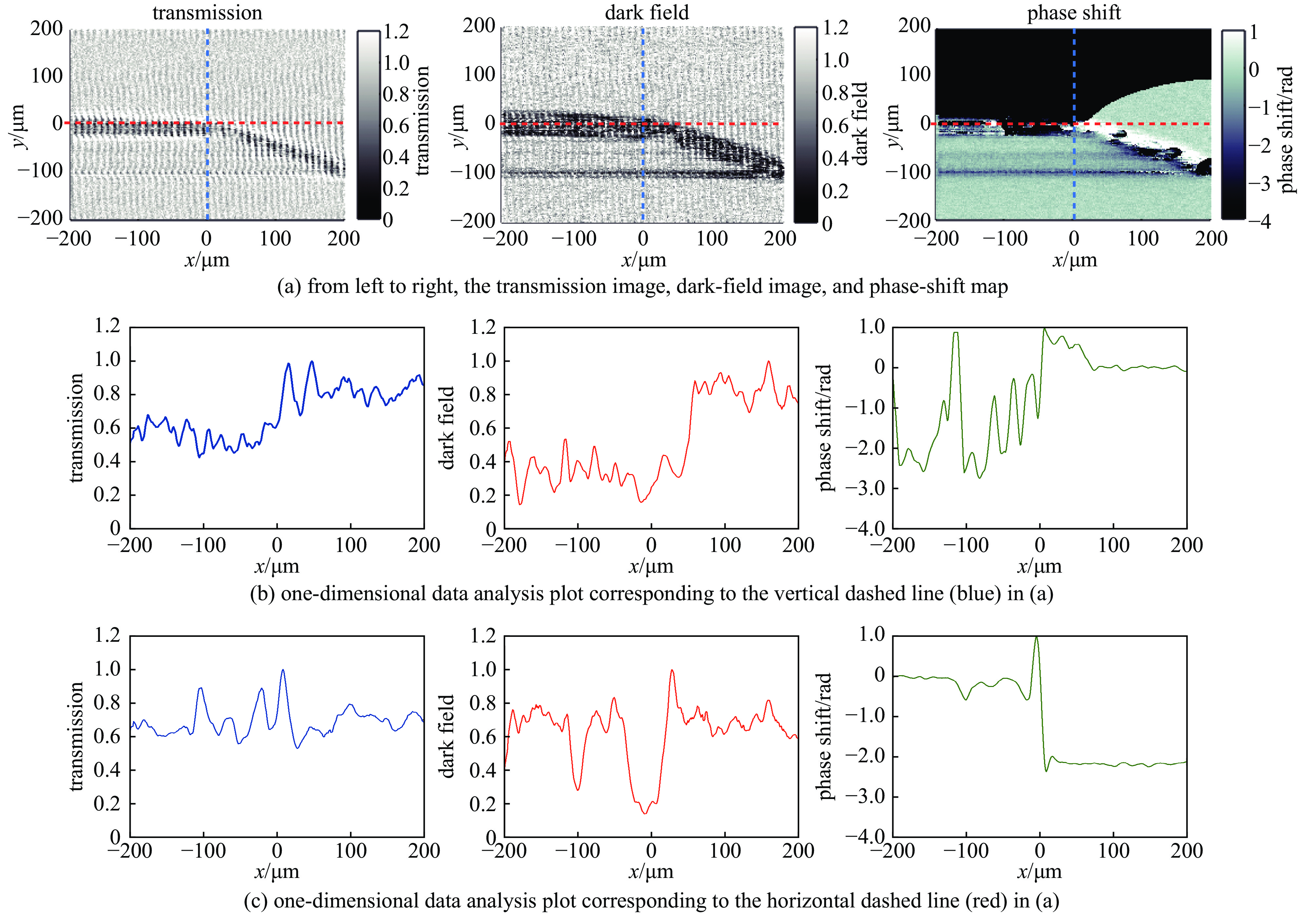
图(15)
计量
- 文章访问数: 667
- HTML全文浏览量: 684
- PDF下载量: 86
- 被引次数: 0
